Copyright
©The Author(s) 2025.
World J Clin Oncol. Mar 24, 2025; 16(3): 101236
Published online Mar 24, 2025. doi: 10.5306/wjco.v16.i3.101236
Published online Mar 24, 2025. doi: 10.5306/wjco.v16.i3.101236
Figure 1 Liver magnetic resonance imaging enhanced findings.
Showing a 20 mm × 18 mm nodule in the hepatic segment VIII with A: Long T1; B: Long T2; and C: Hyperintensity in diffusion-weighted imaging.
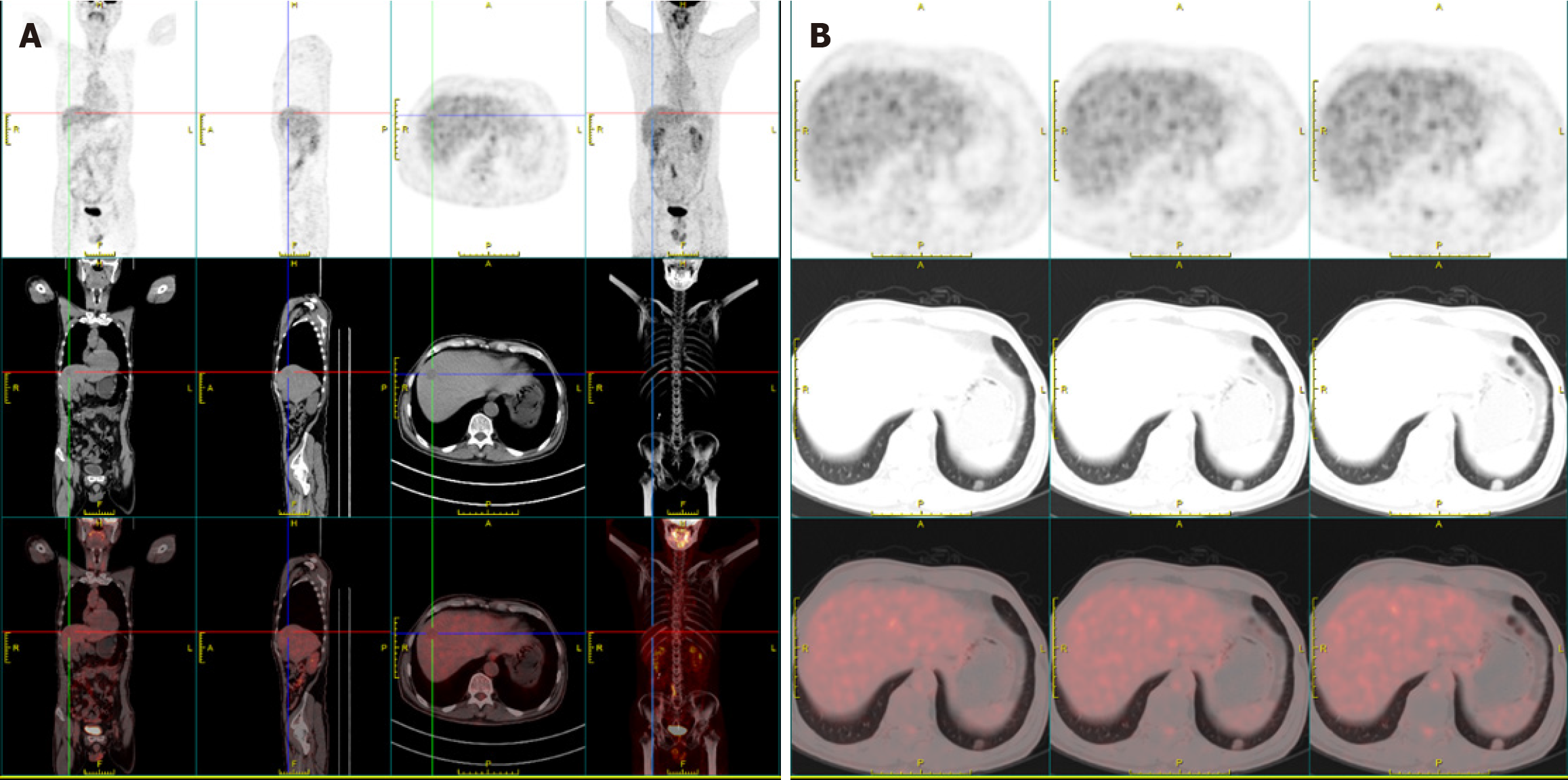
Figure 2 Positron emission tomography-computed tomography findings.
A: Hepatic segment VIII hypodense nodule with a low glucose metabolism; B: Pulmonary nodule with slightly elevated glucose metabolism.
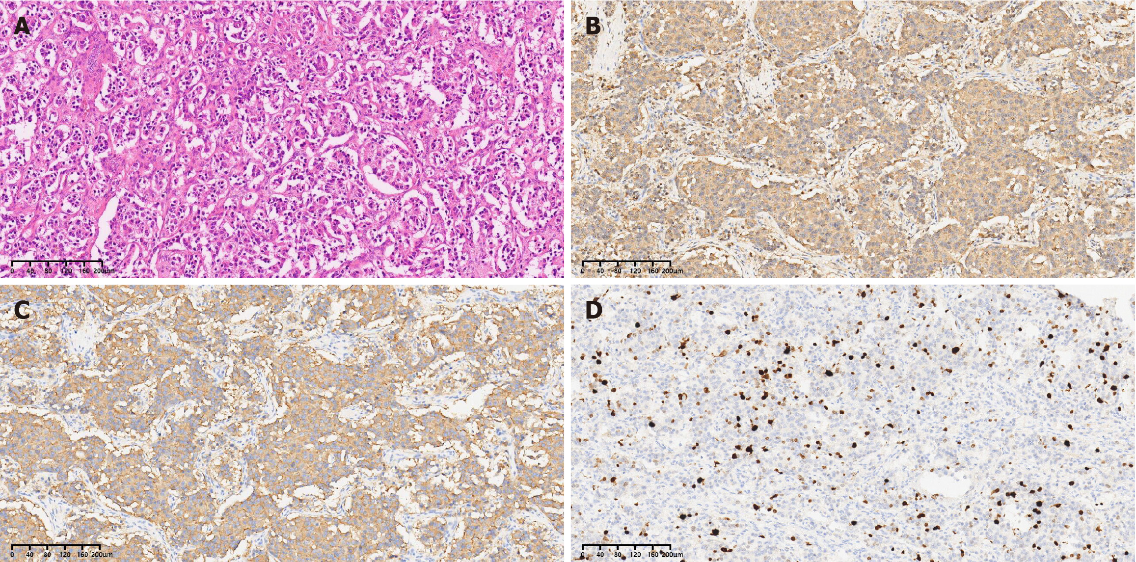
Figure 3 Microscopic findings.
A: Hematoxylin and eosin staining; B: Synaptophysin immunohistochemistry; C: Chromogranin A immunohistochemistry; D: Ki67 immunohistochemistry.
- Citation: Lv HY, Liu MX, Hong WT, Li XW. Primary hepatic neuroendocrine tumor with a suspicious pulmonary nodule: A case report and literature review. World J Clin Oncol 2025; 16(3): 101236
- URL: https://www.wjgnet.com/2218-4333/full/v16/i3/101236.htm
- DOI: https://dx.doi.org/10.5306/wjco.v16.i3.101236


