Copyright
©The Author(s) 2025.
World J Clin Oncol. Feb 24, 2025; 16(2): 99635
Published online Feb 24, 2025. doi: 10.5306/wjco.v16.i2.99635
Published online Feb 24, 2025. doi: 10.5306/wjco.v16.i2.99635
Figure 1 Endoscopic plus mass biopsy and postoperative pathology of a circumferential bulging mass in the rectum.
A: Endoscopic image of a malignant rectal tumor; B: Hematoxylin-eosin staining (× 100) of rectal mass biopsy tissue; C: Hematoxylin-eosin staining (× 100) of postoperative rectal mass tissue.
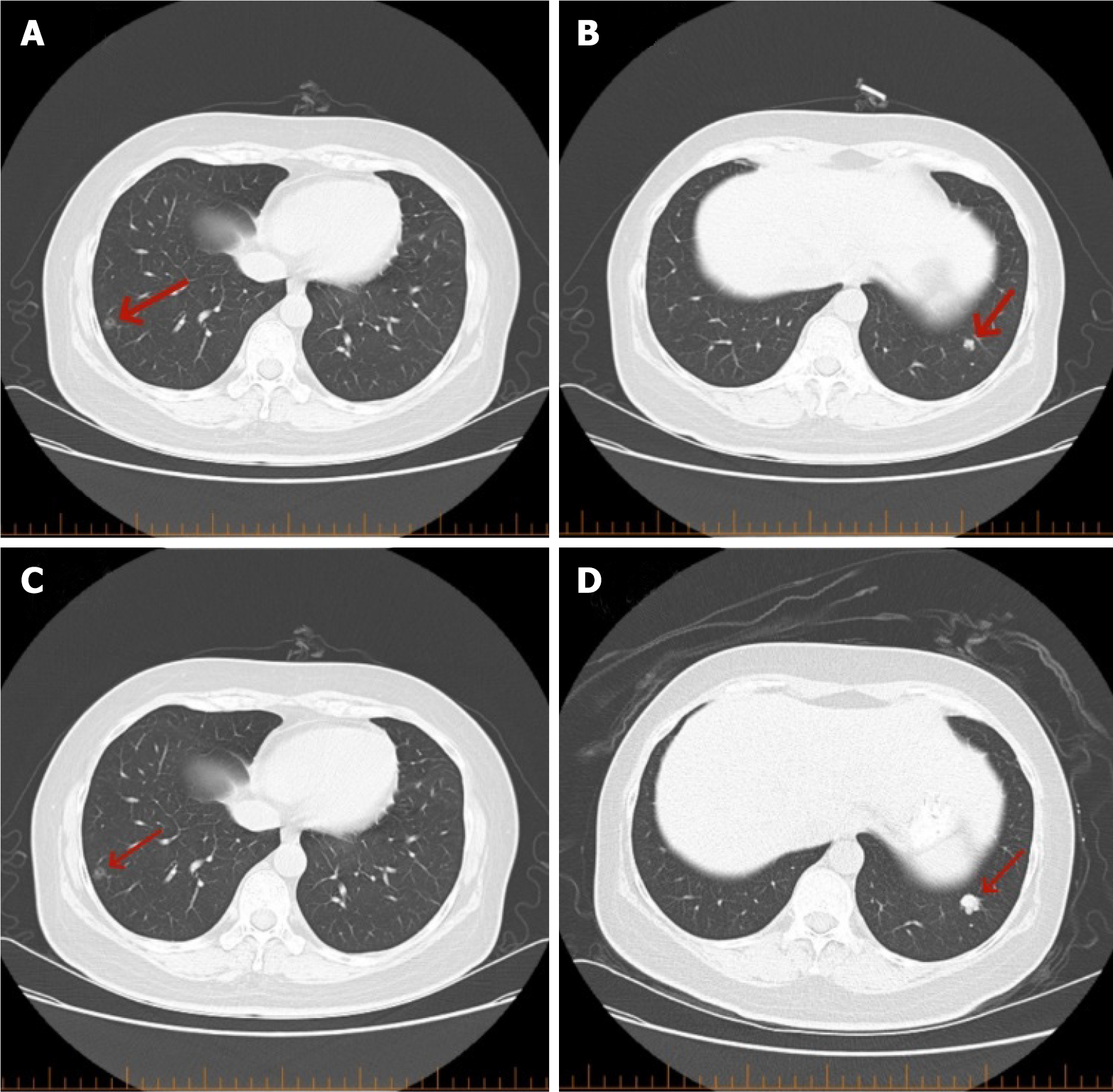
Figure 2 Whole-body computed tomography scan.
A: A ground-glass nodule (red arrow) is visible in the outer basal segment of the right lower lobe on the computed tomography scan taken on July 10, 2021; B: A nodule (red arrow) is observed in the outer basal segment of the left lower lobe; C: A ground-glass nodule (red arrow) is visible in the outer basal segment of the right lower lobe on the computed tomography scan taken on January 25, 2022; D: A nodule (red arrow) is observed in the outer basal segment of the left lower lobe on the computed tomography scan taken on January 25, 2022.
Figure 3 Hematoxylin-eosin staining and immunohistochemistry of a tumor in the lower lobe of the left lung.
A: Hematoxylin-eosin staining of lung biopsy tissue (× 100); B: CK20-immunostained positive tumor cells (× 100); C: CDX2-immunostained positive tumor cells (× 100).
Figure 4 Hematoxylin-eosin staining and immunohistochemical analysis of the upper and lower lobes of the right lung.
A: Hematoxylin-eosin staining of lung biopsy tissue (×40); B: Hematoxylin-eosin staining of lung biopsy tissue (× 100); C: TTF-1-immunostained positive tumor cells (× 100).
- Citation: Zhou FY, Song FH, Cheng ZH, Wu S. Discovery of primary lung cancer following resection of rectal cancer lung metastasis: A case report. World J Clin Oncol 2025; 16(2): 99635
- URL: https://www.wjgnet.com/2218-4333/full/v16/i2/99635.htm
- DOI: https://dx.doi.org/10.5306/wjco.v16.i2.99635


