Copyright
©The Author(s) 2025.
World J Clin Oncol. Feb 24, 2025; 16(2): 98223
Published online Feb 24, 2025. doi: 10.5306/wjco.v16.i2.98223
Published online Feb 24, 2025. doi: 10.5306/wjco.v16.i2.98223
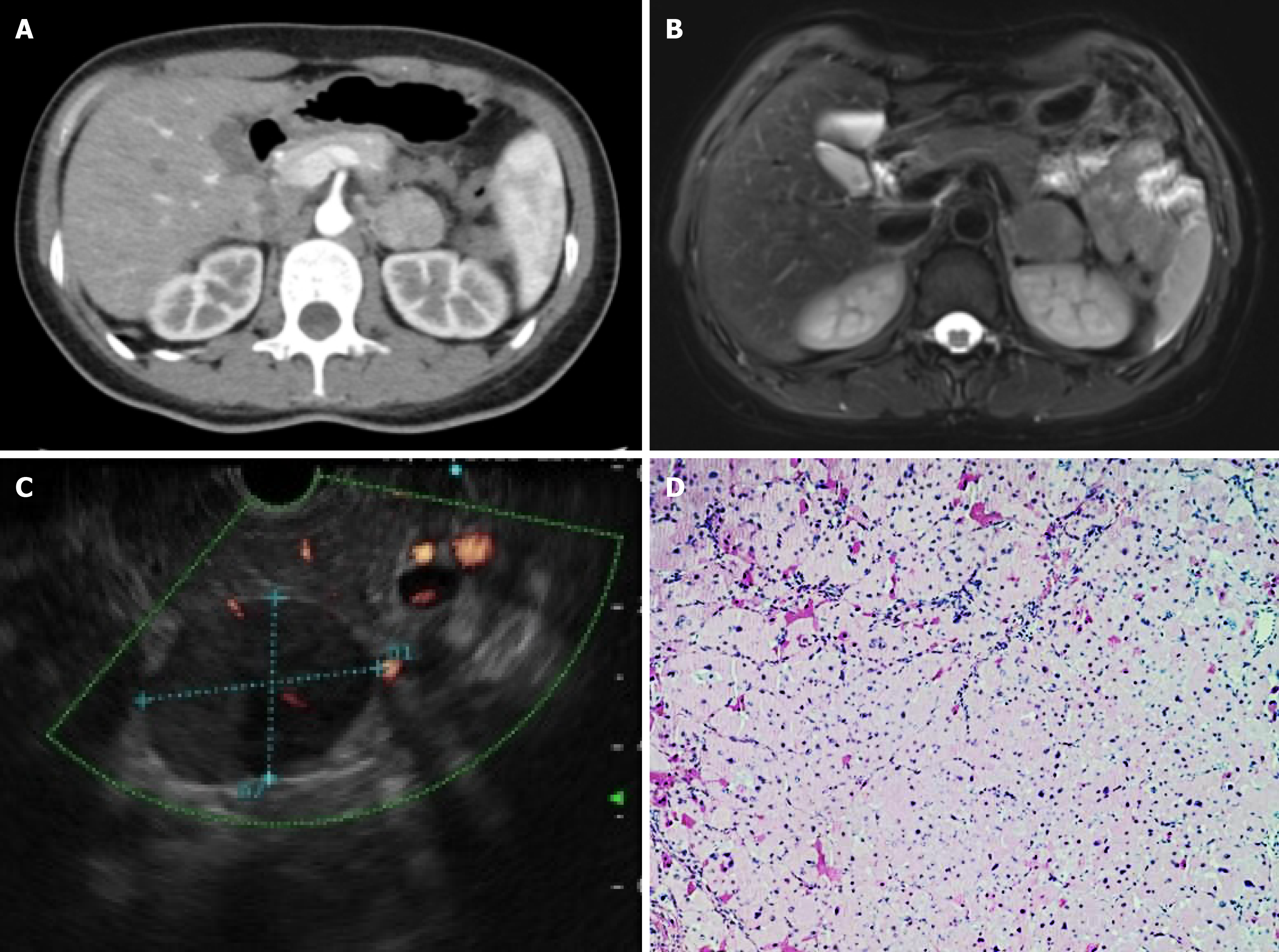
Figure 1 Imaging and histopathological pictures.
A: Computed tomography with contrast shows a heterogeneously enhanced mass; B: Magnetic resonance with contrast showing the circumferential enhancement of the lesion with clear border; C: Endoscopic ultrasound shows a heterogeneous hypoechoic mass behind the body-caudal junction of the pancreas; D: Histopathology section showing diffuse proliferation of oncocytes (× 20).
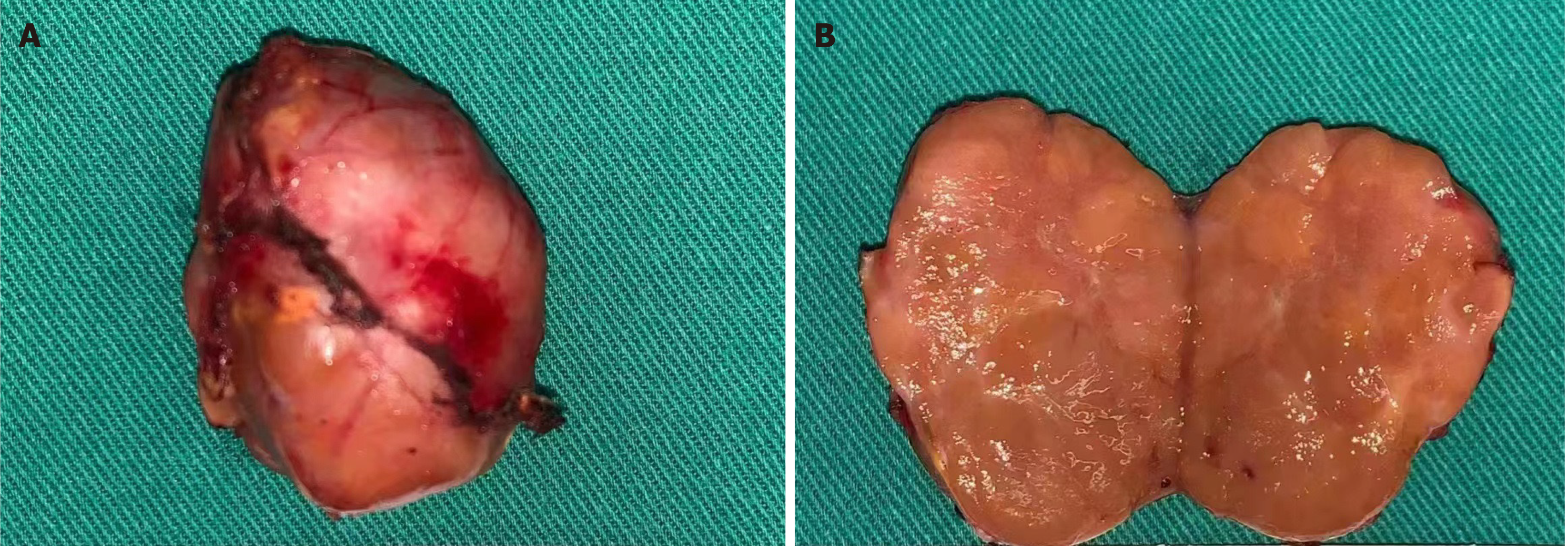
Figure 2 Postoperative image of the resected tumor.
A: The tumor (size 4 cm × 3 cm × 3 cm) was nodular with intact envelope; B: The yellow-brown cut surface.
- Citation: Chen H, Jing X. Individualized treatment guided by endoscopic ultrasound-guided fine-needle aspiration for adrenocortical oncocytoma: A case report. World J Clin Oncol 2025; 16(2): 98223
- URL: https://www.wjgnet.com/2218-4333/full/v16/i2/98223.htm
- DOI: https://dx.doi.org/10.5306/wjco.v16.i2.98223


