Copyright
©The Author(s) 2025.
World J Clin Oncol. Feb 24, 2025; 16(2): 97296
Published online Feb 24, 2025. doi: 10.5306/wjco.v16.i2.97296
Published online Feb 24, 2025. doi: 10.5306/wjco.v16.i2.97296
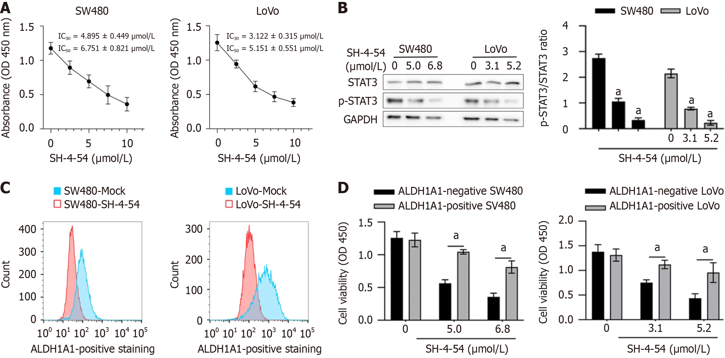
Figure 1 The effects of SH-4-45 on ALDH1A1-positive proportion of colorectal cancer cells.
A: After being cultured with 1-10 μmol/L of SH-4-45 for 24 hours, cell viability was measured by performing CCK-8 assay; B: Western blot was performed to detect STAT3 phosphorylation; C: Flow cytometry assay was performed to detect ALDH1A1-positive proportion; D: After being cultured in SH-4-54 for 24 hours, cell viability was detected. CSCs: Cancer stem cells.
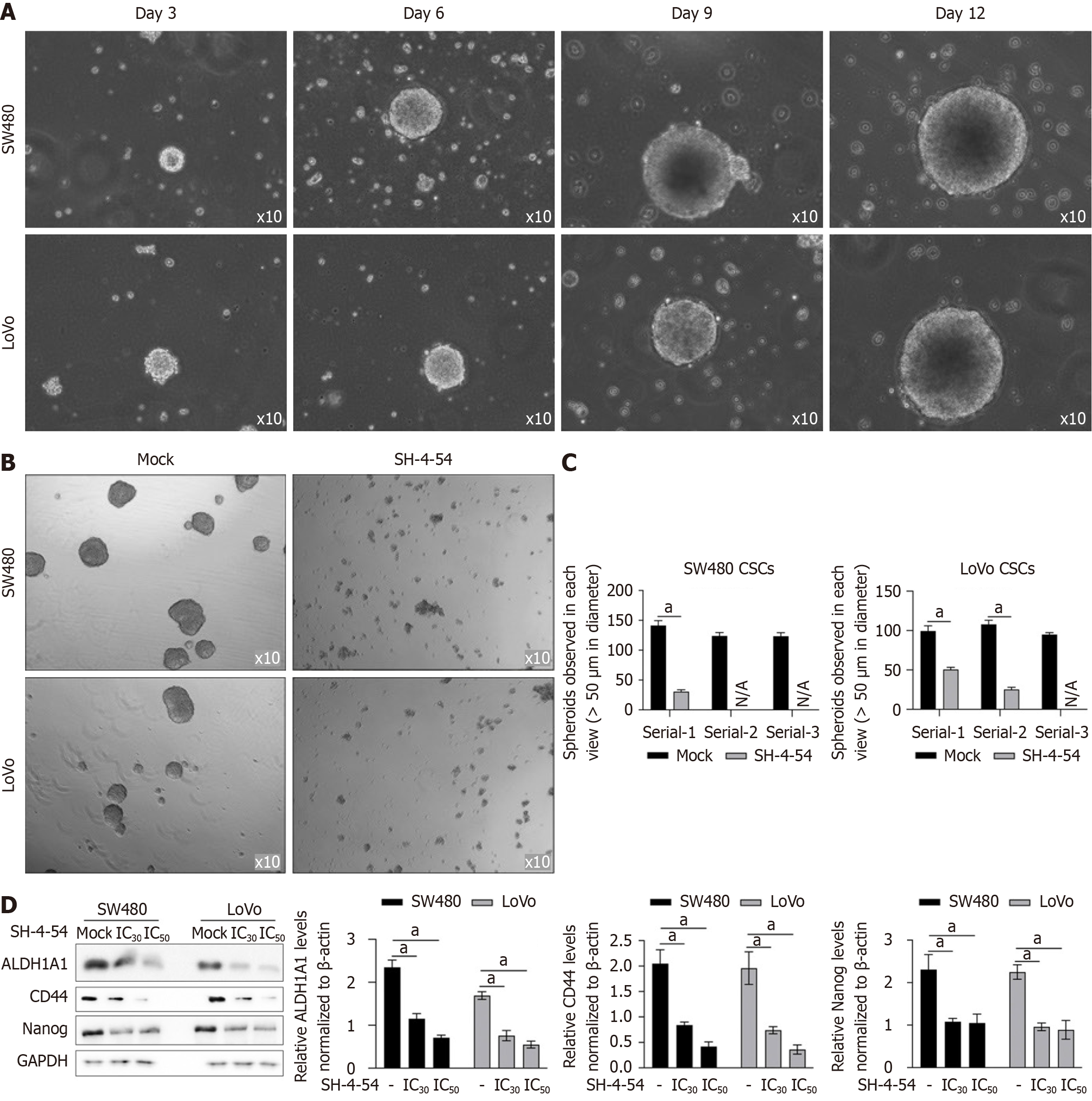
Figure 2 SH-4-54 inhibited stemness in cancer stem cells.
A-C: SW480 or LoVo cells were suspended and cultured in serum-free medium and kept for 12-days. At day 3, 6, 9, and 12, spheroids were imaged. After addition of SH-4-54 for 72 hours, spheroids were imaged (B) and self-renewal capacity was analyzed by performing serial replating assay (C); D: After SH-4-54 treatment, ALDH1A1, CD44 and Nanog were detected by performing western blot. After addition of SH-4-54 for 72 hours, cells were collected and stemness hallmarkers were detected by western blot, including CD44, Nanog, and ALDH1A1. aP < 0.05 vs mock group. PCs: Parental cells; CSCs: Cancer stem cells.
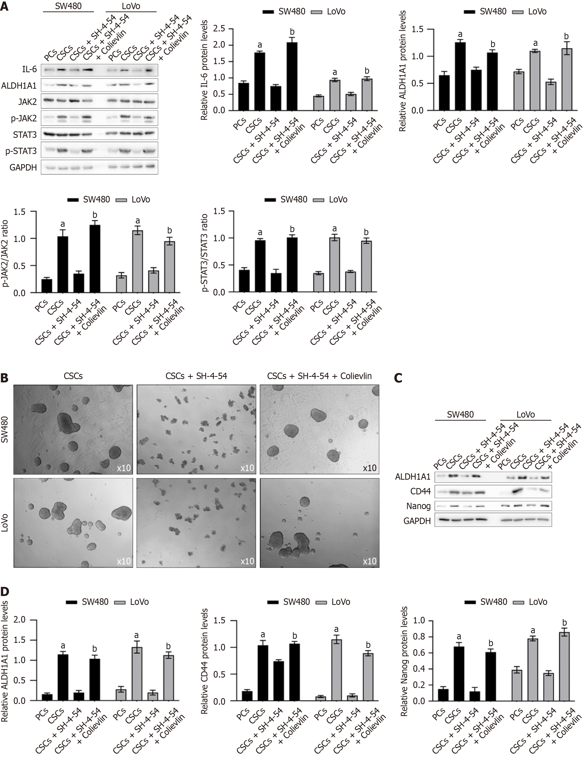
Figure 3 SH-4-54 decreased stemness hallmarkers via inhibiting phosphorylation of STAT3.
A: Cancer stem cells (CSCs) were cultured with SH-4-54 with addition Colievlin or not, for 24 hours, then IL-6, JAK2/STAT3 signaling and stemness hallmarkers were detected by performing Western blot; B: With the presence of SH-4-54 with or without Colievlin, spheroid formation was imaged after 12-day incubation; C: After addition of Colievlin, the stemness hallmarkers were detected by performing western blot, including ALDH1A1, CD44 and Nanog. aP < 0.05 vs CSCs group; bP < 0.05 vs CSCs+SH-4-54 group. PCs: Parental cells; CSCs: Cancer stem cells.
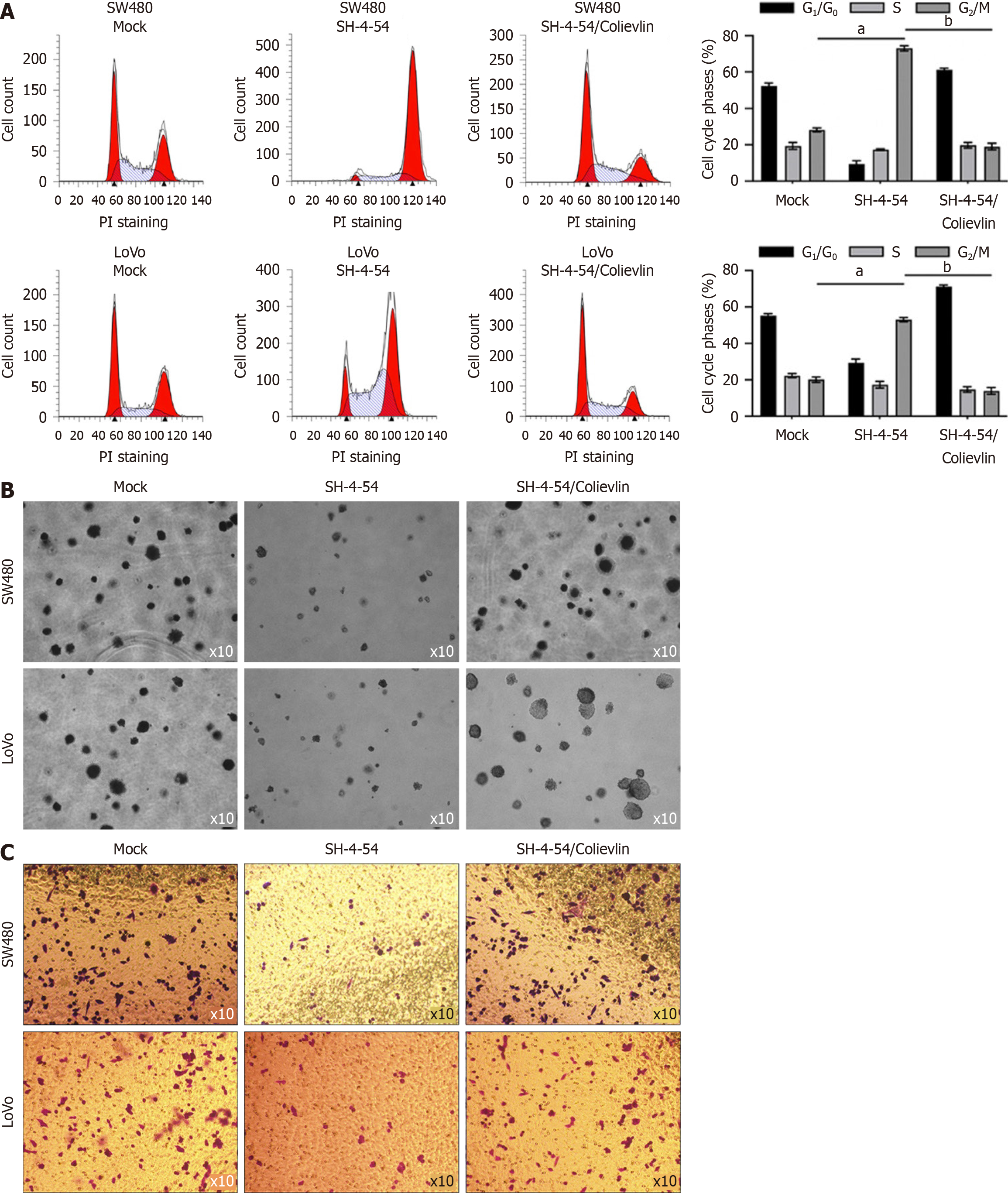
Figure 4 SH-4-54 treatment decreased malignancies in cancer stem cells derived from SW480 and LoVo.
A: After being cultured in IC30 concentration of SH-4-54 for 24 hours, cells were fixed by 4% paraformaldehyde and analyzed by performing flow cytometry to detect DNA content; B: After being co-cultured in soft agar medium containing IC30 concentration of SH-4-54 for 12 days, formed colonies were imaged; C: After being cultured in IC30 concentration of SH-4-54 for 48 hours, Transwell assay was performed and transferred cells to lower membrane in five random views were calculated. aP < 0.05 vs PCs group; bP < 0.05 vs cancer stem cells group.
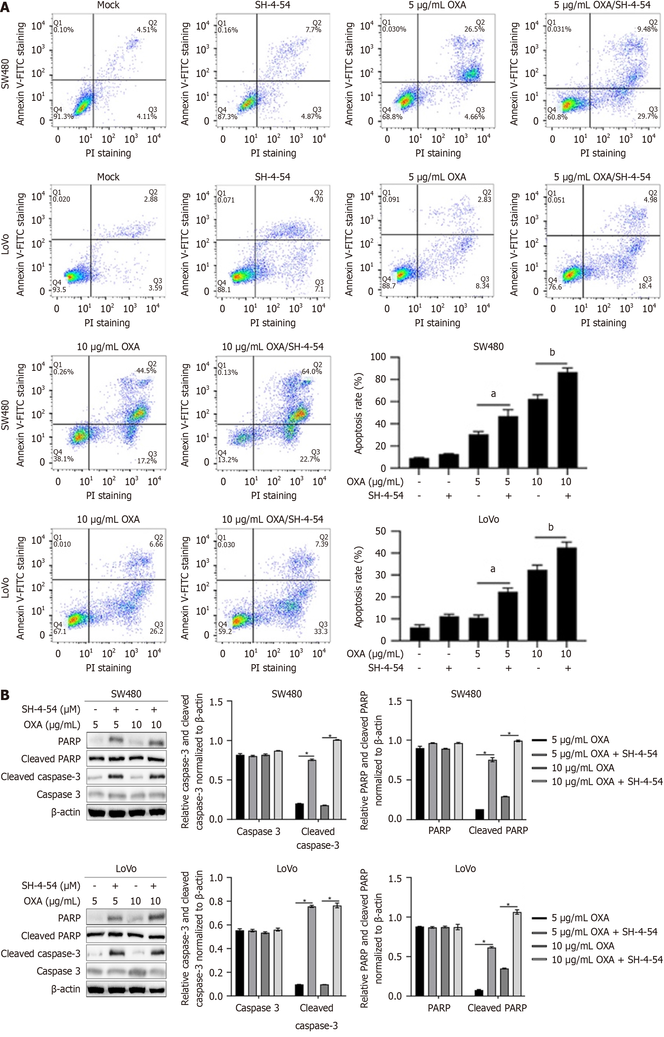
Figure 5 SH-4-54 treatment sensitizes cancer stem cells against oxaplatin.
A: After being culture with 5 or 10 μg/mL of oxaplatin (OXA), SH-4-54 was added for co-culture, and then cell apoptosis was analyzed by performing Annexin V-FITC/PI double staining, followed by a flow cytometry assay; B: Total protein was fractioned and performed to detect apoptotic-related hallmarkers, including PARP, cleaved-PARP, caspase 3, and cleaved caspase-3. aP < 0.05 vs 5 μg/mL of OXA group; bP < 0.05 vs 10 μg/mL of OXA group. OXA: Oxaplatin.
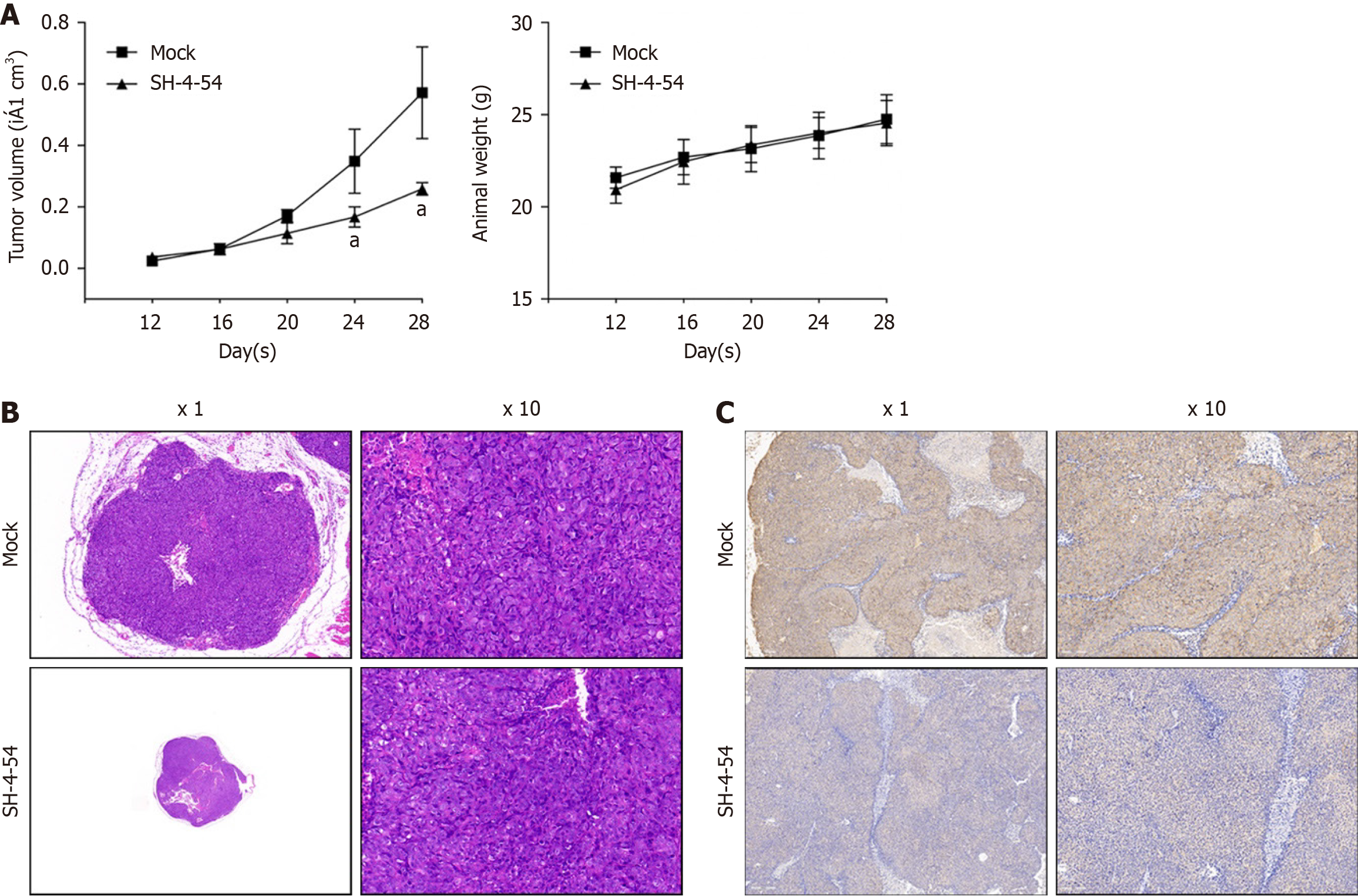
Figure 6 SH-4-54 inhibited tumor growth of cancer stem cells in vivo.
A: 5 × 10⁵ cancer stem cells derived from SW480 was planted subcutaneously in nude mice and maintained for 30 days. Tumor growth curve (left panel) and animal weight curve (right panel) was drawn; B and C: Tumor tissues were fixed and sectioned for H&E staining (B) and Ki67 staining (C). aP < 0.05 vs mock group.
- Citation: Zhang XF, Chen Q, Jiang Q, Hu QY. Targeting STAT3 with SH-4-54 suppresses stemness and chemoresistance in cancer stem-like cells derived from colorectal cancer. World J Clin Oncol 2025; 16(2): 97296
- URL: https://www.wjgnet.com/2218-4333/full/v16/i2/97296.htm
- DOI: https://dx.doi.org/10.5306/wjco.v16.i2.97296


