Copyright
©The Author(s) 2025.
World J Clin Oncol. Feb 24, 2025; 16(2): 97248
Published online Feb 24, 2025. doi: 10.5306/wjco.v16.i2.97248
Published online Feb 24, 2025. doi: 10.5306/wjco.v16.i2.97248
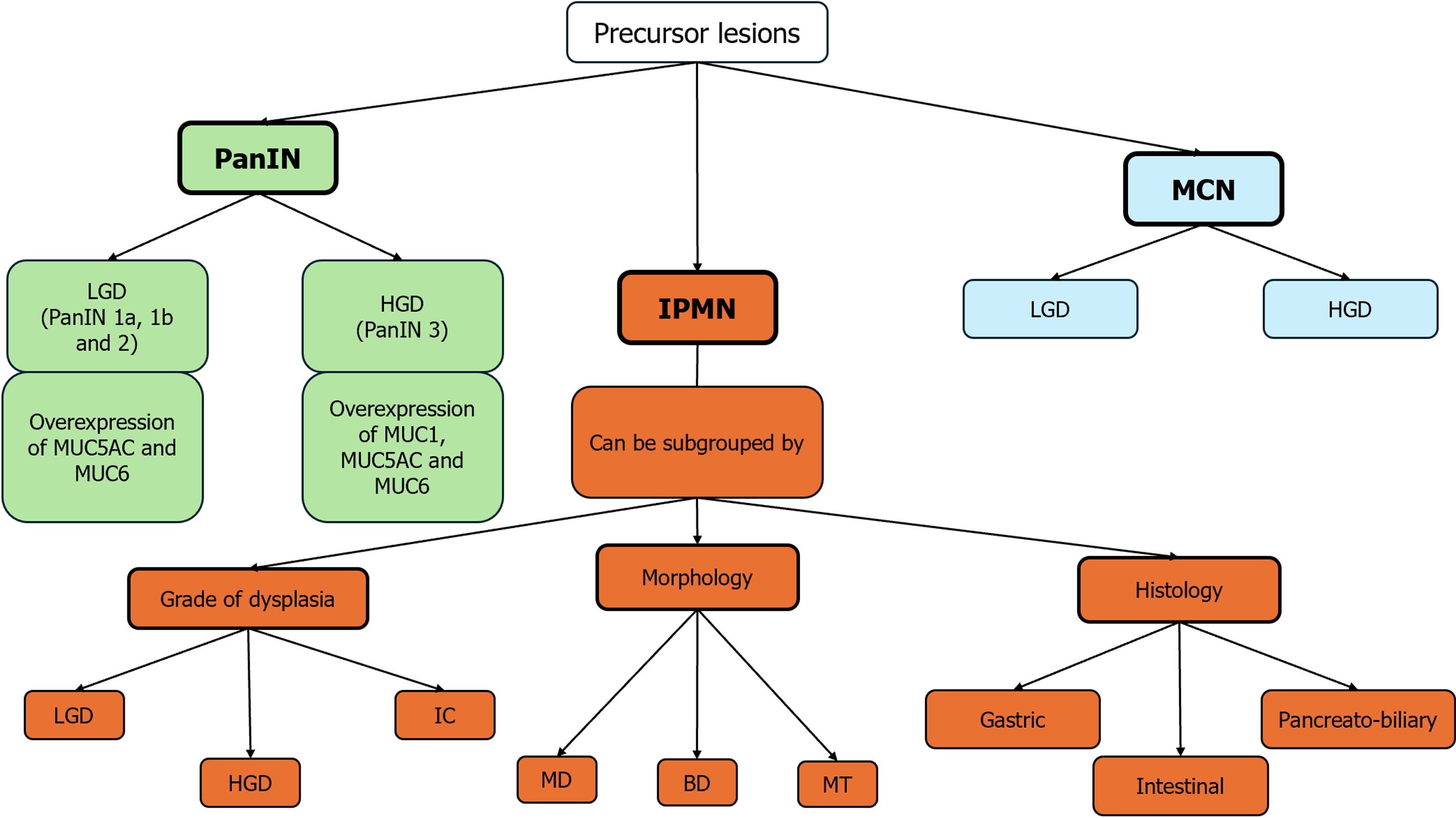
Figure 1 Precursor lesions of pancreatic cancer: A schematic overlook.
BD: Branch duct; HGD: High grade dysplasia; IC: Invasive cancer; IPMN: Intraductal papillary mucinous neoplasm; LGD: Low grade dysplasia; MCN: Mucinous cystic neoplasm; MD: Main duct; MT: Mixed type; PanIN: Pancreatic intraepithelial neoplasia.
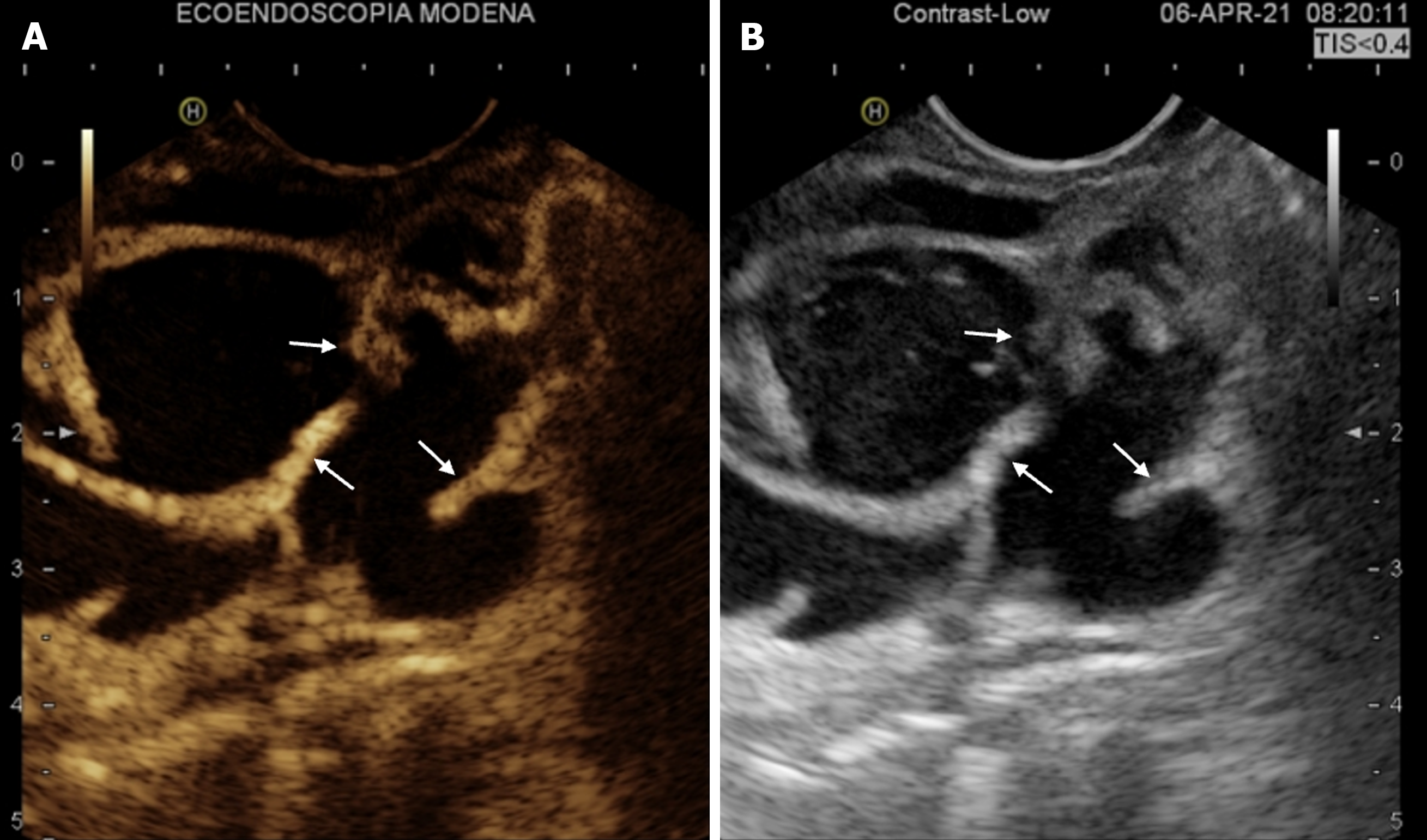
Figure 2 Contrast-enhanced endoscopic ultrasound study of intraductal papillary mucinous neoplasms-branch duct.
A: Contrast-enhanced-endoscopic ultrasound (EUS) of a large multilocular cyst with contrast-enhanced thickened septa (arrows); B: EUS B-mode image of the same lesion.
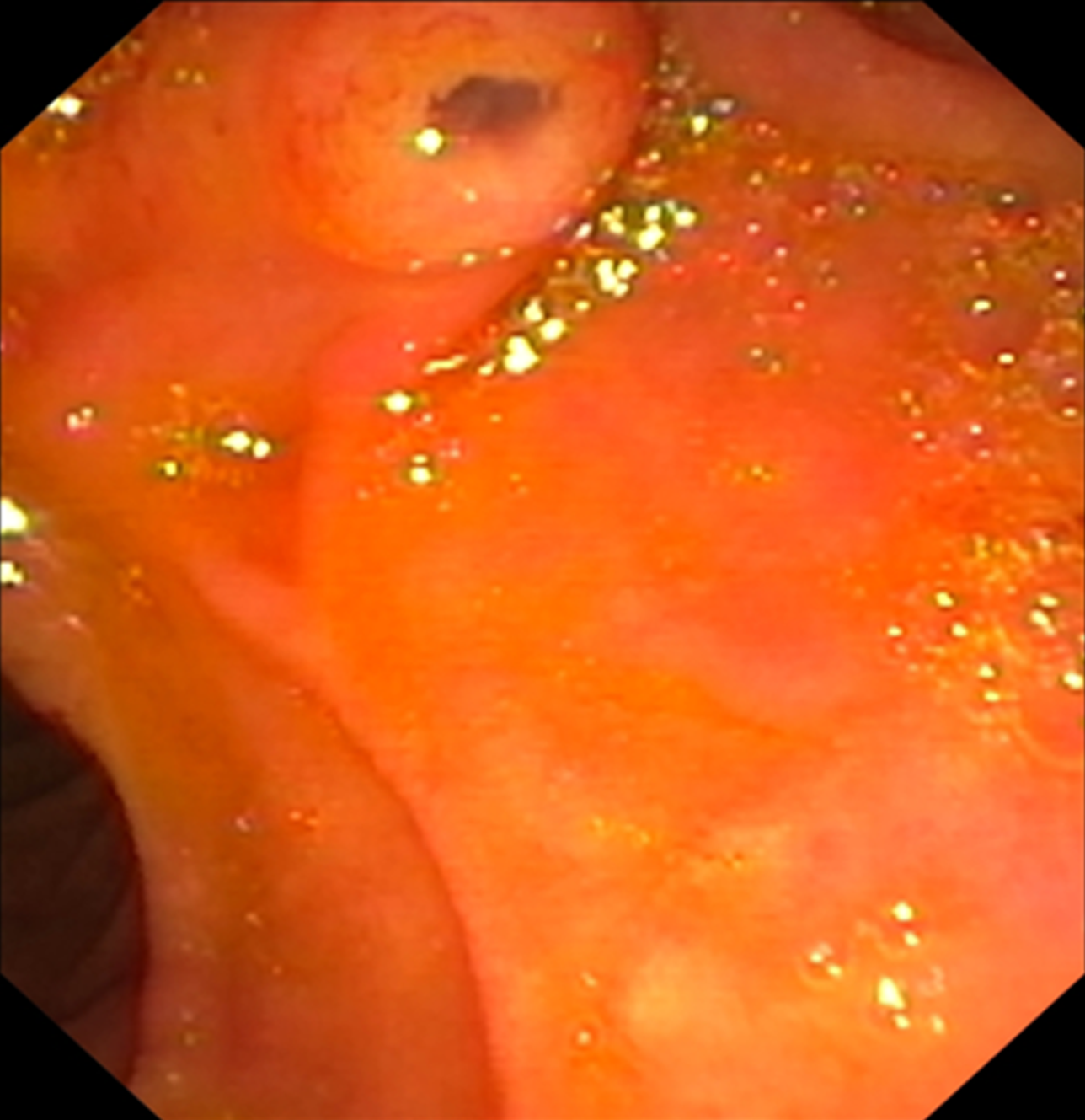
Figure 3 Fish-eye sign.
Endoscopic view of a bulging ampulla actively extruding mucus.
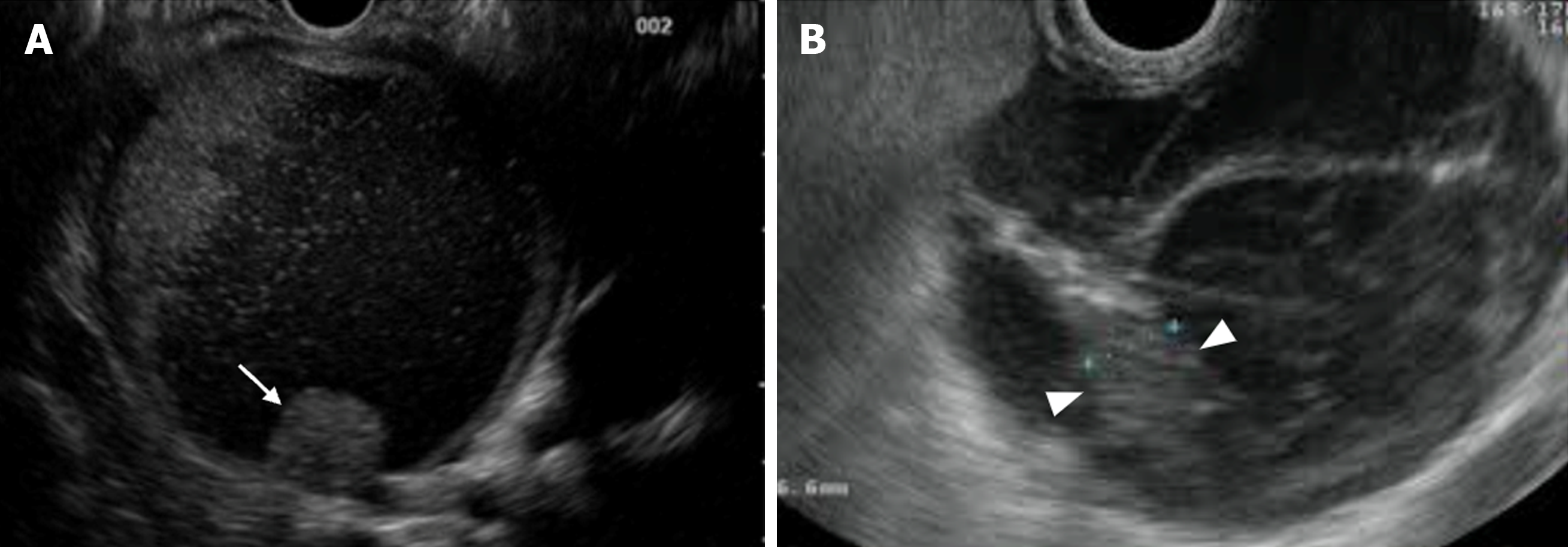
Figure 4 Mucinous cystic neoplasms with “worrisome” features.
A: Mural nodule (arrow); B: Thickened septum (arrow heads).
- Citation: Cocca S, Pontillo G, Lupo M, Lieto R, Marocchi M, Marsico M, Dell'Aquila E, Mangiafico S, Grande G, Conigliaro R, Bertani H. Pancreatic cancer: Future challenges and new perspectives for an early diagnosis. World J Clin Oncol 2025; 16(2): 97248
- URL: https://www.wjgnet.com/2218-4333/full/v16/i2/97248.htm
- DOI: https://dx.doi.org/10.5306/wjco.v16.i2.97248


