Copyright
©The Author(s) 2024.
World J Clin Oncol. Sep 24, 2024; 15(9): 1232-1238
Published online Sep 24, 2024. doi: 10.5306/wjco.v15.i9.1232
Published online Sep 24, 2024. doi: 10.5306/wjco.v15.i9.1232
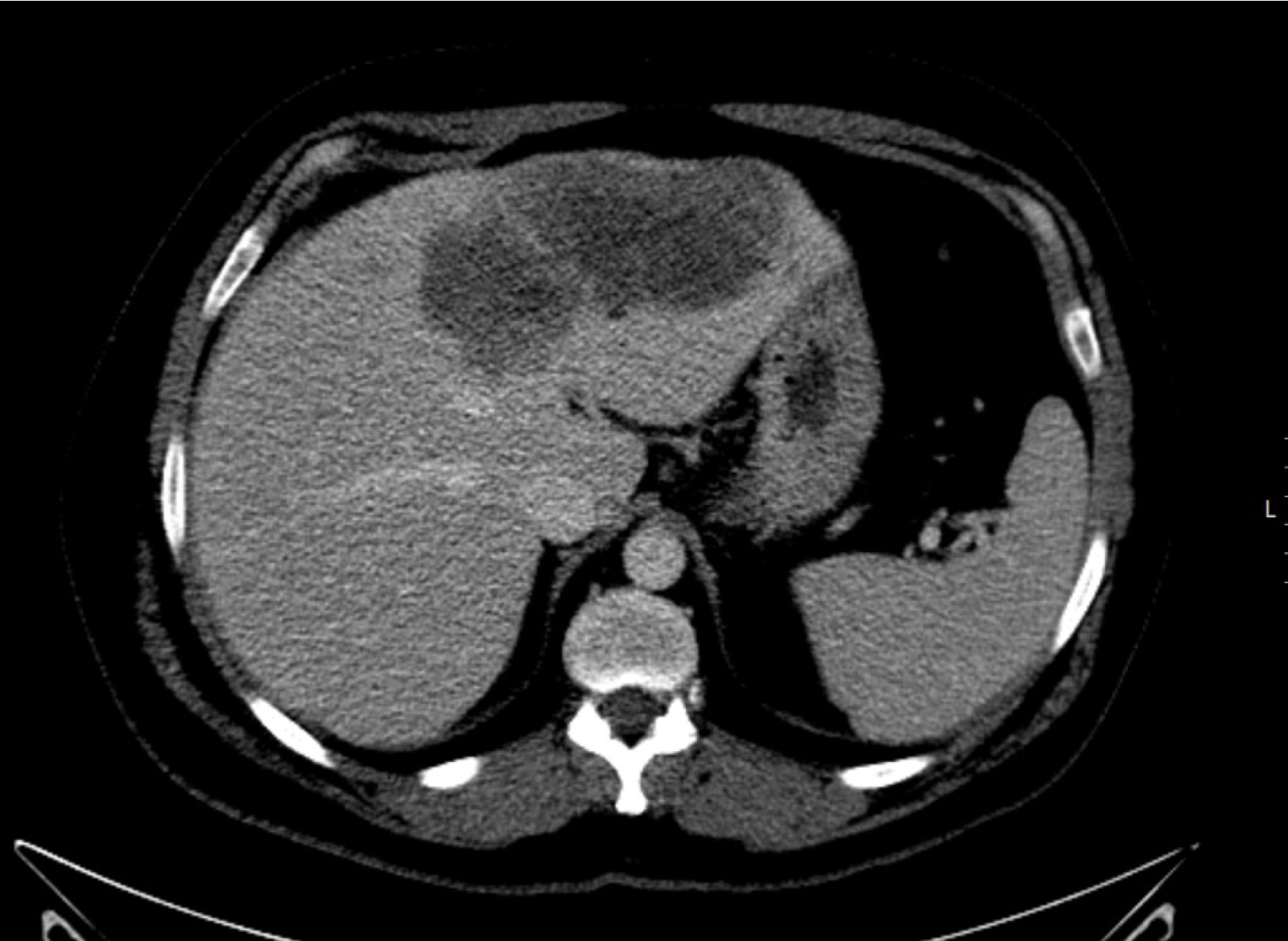
Figure 1
Computed tomography image of the primary nodular lesion in the left hepatic lobe.
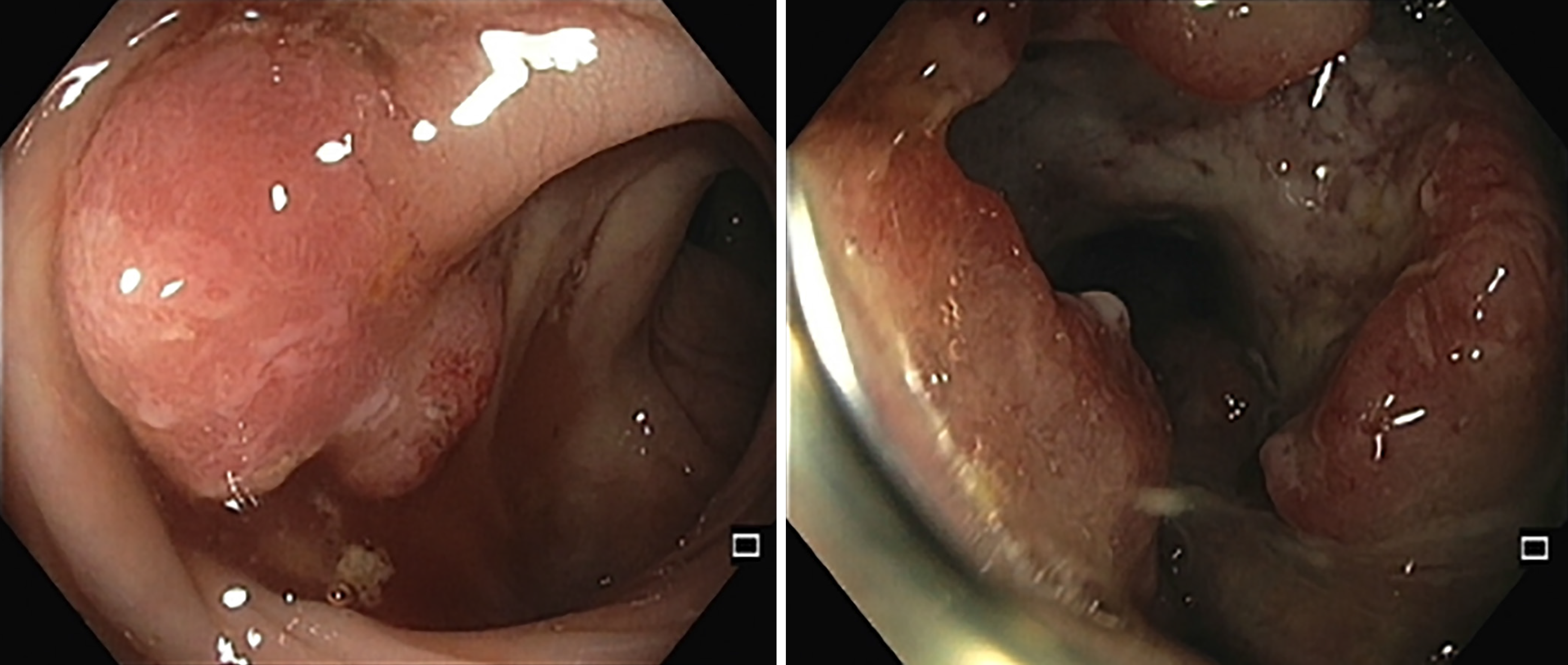
Figure 2
Colonoscopy images of the neoformative lesion at diagnosis.
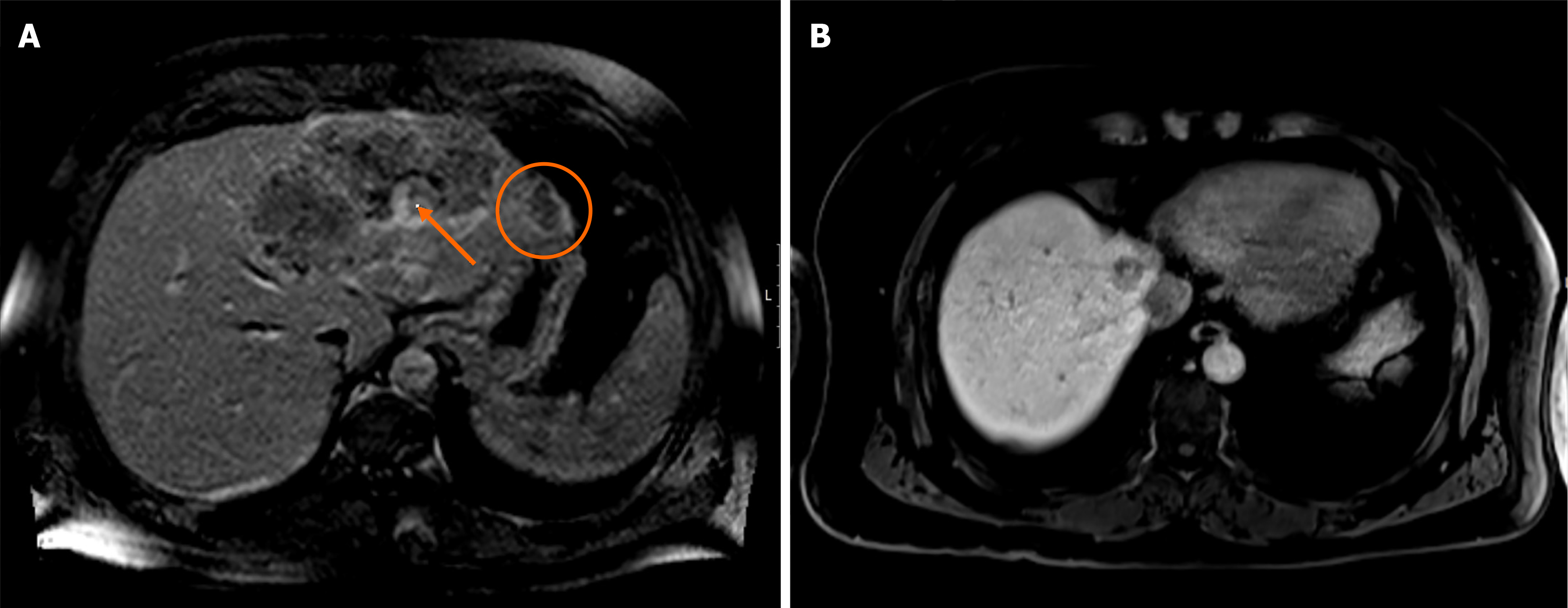
Figure 3 Hepatic magnetic resonance image.
A: Hepatic magnetic resonance image of several confluent hepatic lesions in the left lobe; B: Hepatic magnetic resonance image of one de novo hepatic metastasis of segment VIII and adenopathy next to the caudate lobe.
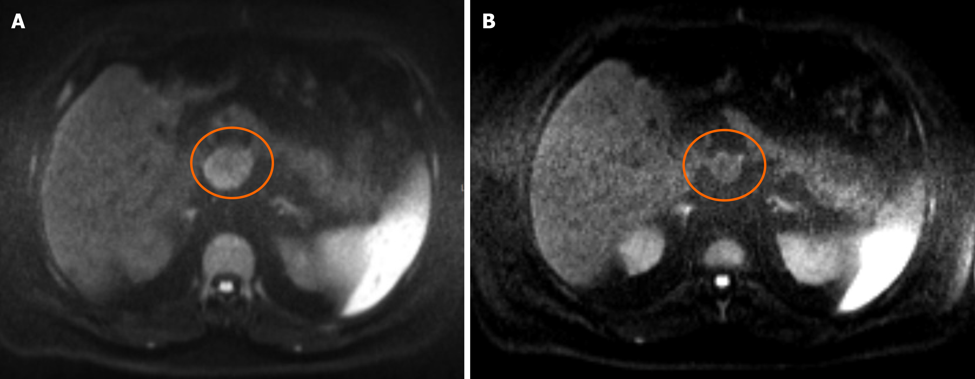
Figure 4 Hepatic magnetic resonance before third-line therapy and after seven cycles of treatment with FOLFIRI and cetuximab.
A: Hepatic magnetic resonance before third-line therapy; B: Hepatic magnetic resonance after seven cycles of treatment with FOLFIRI and cetuximab.
- Citation: Guedes A, Silva S, Custódio S, Capela A. Successful cetuximab rechallenge in metastatic colorectal cancer: A case report. World J Clin Oncol 2024; 15(9): 1232-1238
- URL: https://www.wjgnet.com/2218-4333/full/v15/i9/1232.htm
- DOI: https://dx.doi.org/10.5306/wjco.v15.i9.1232


