Copyright
©The Author(s) 2024.
World J Clin Oncol. Sep 24, 2024; 15(9): 1222-1231
Published online Sep 24, 2024. doi: 10.5306/wjco.v15.i9.1222
Published online Sep 24, 2024. doi: 10.5306/wjco.v15.i9.1222
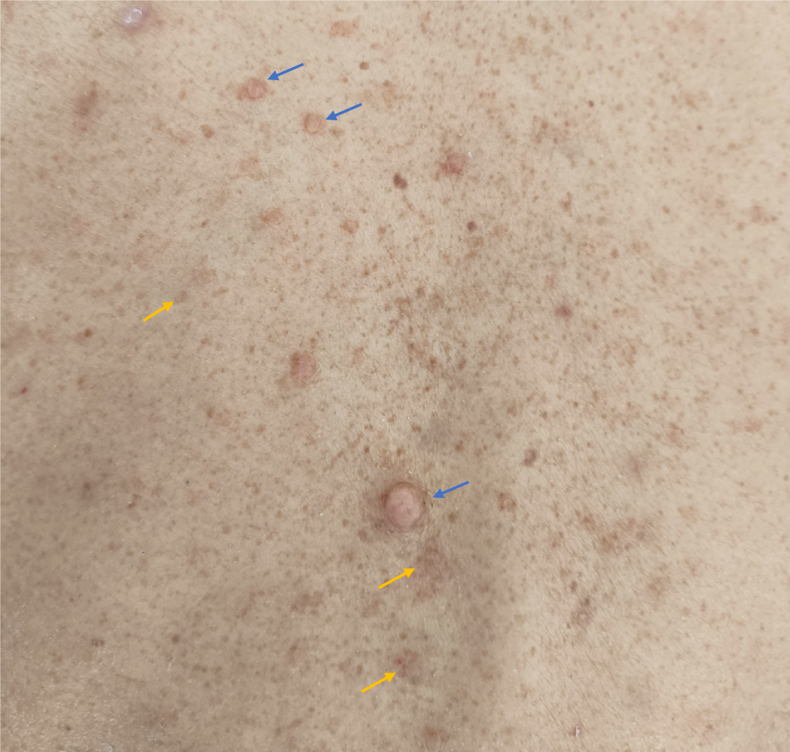
Figure 1 A view of the skin on the patient’s back.
Numerous café-au-lait macules (the yellow arrows) and cutaneous neurofibromas (the blue arrows) can be seen.
Figure 2 Findings of the abdominal contrast-enhanced computed tomography scan.
A: A tumor (the yellow arrow) 1.2 cm × 1.4 cm in the periampullary region, with progressive enhancement; B: Multiple hemangiomas (the yellow arrow) in the S8 segment of the liver; C: Localized thickening of the skin and subcutaneous tissue (the yellow arrow) in the lower anterior abdominal wall.
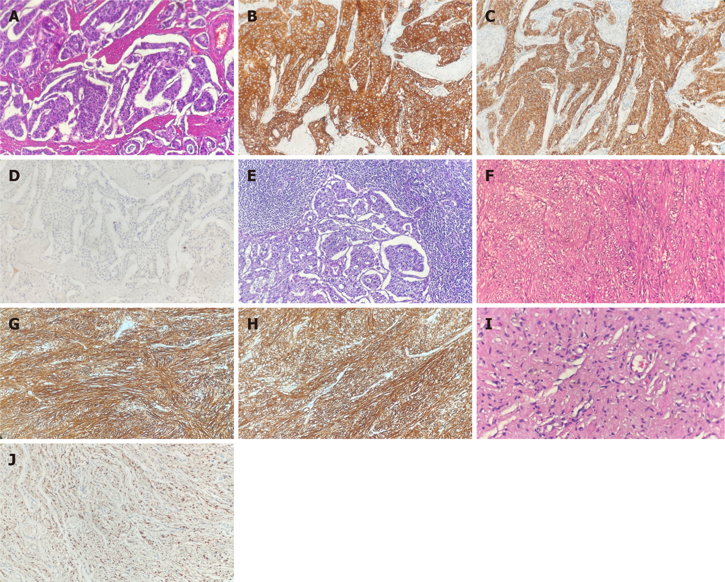
Figure 3 Microscopic findings of the pancreatic tumor.
A: Histologic hematoxylin and eosin (HE) stain of the duodenal neuroendocrine tumor (NET) (magnification × 200); B and C: Expression of synaptophysin (B) and chromogranin A (C) were positive on immunohistochemical stain in the NET; D: Ki67 labeling index of the NET was 1%; E: Metastatic tumors in peripancreatic lymph nodes; F: HE stain of the stromal tumor in the serous surface of the duodenum; G and H: Expression of CD 117(G) and DOG-1 (H) were positive on immunohistochemical stain in the stromal tumor; I: HE stain of the cutaneous neurofibroma; J: Expression of S100 protein was positive in the neurofibroma (magnification × 200).
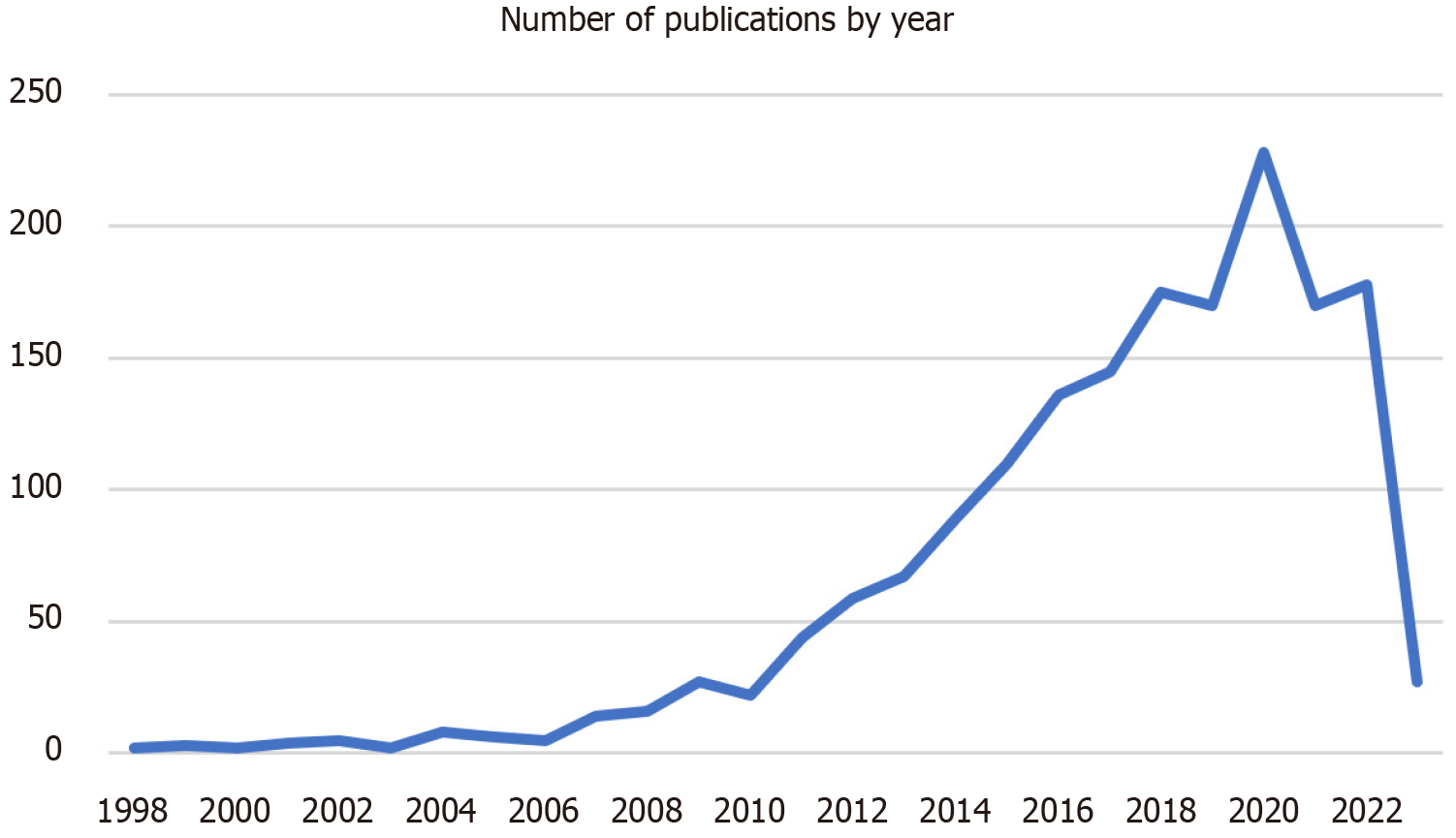
Figure 4 Number of publications by year.
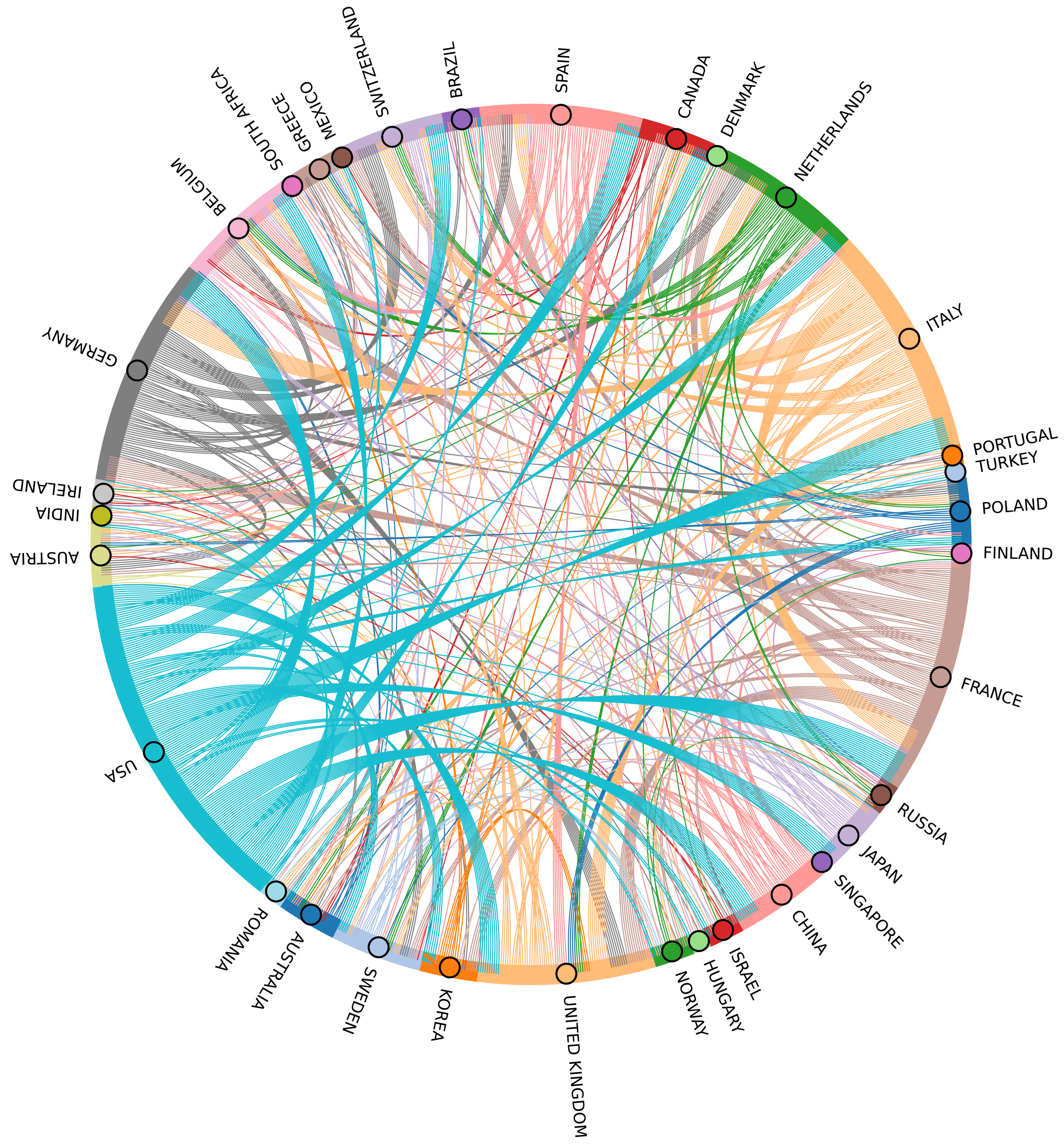
Figure 5 Cooperative relationships between countries/regions.
To make the plot more concise, the countries with frequencies less than 3 were deleted.
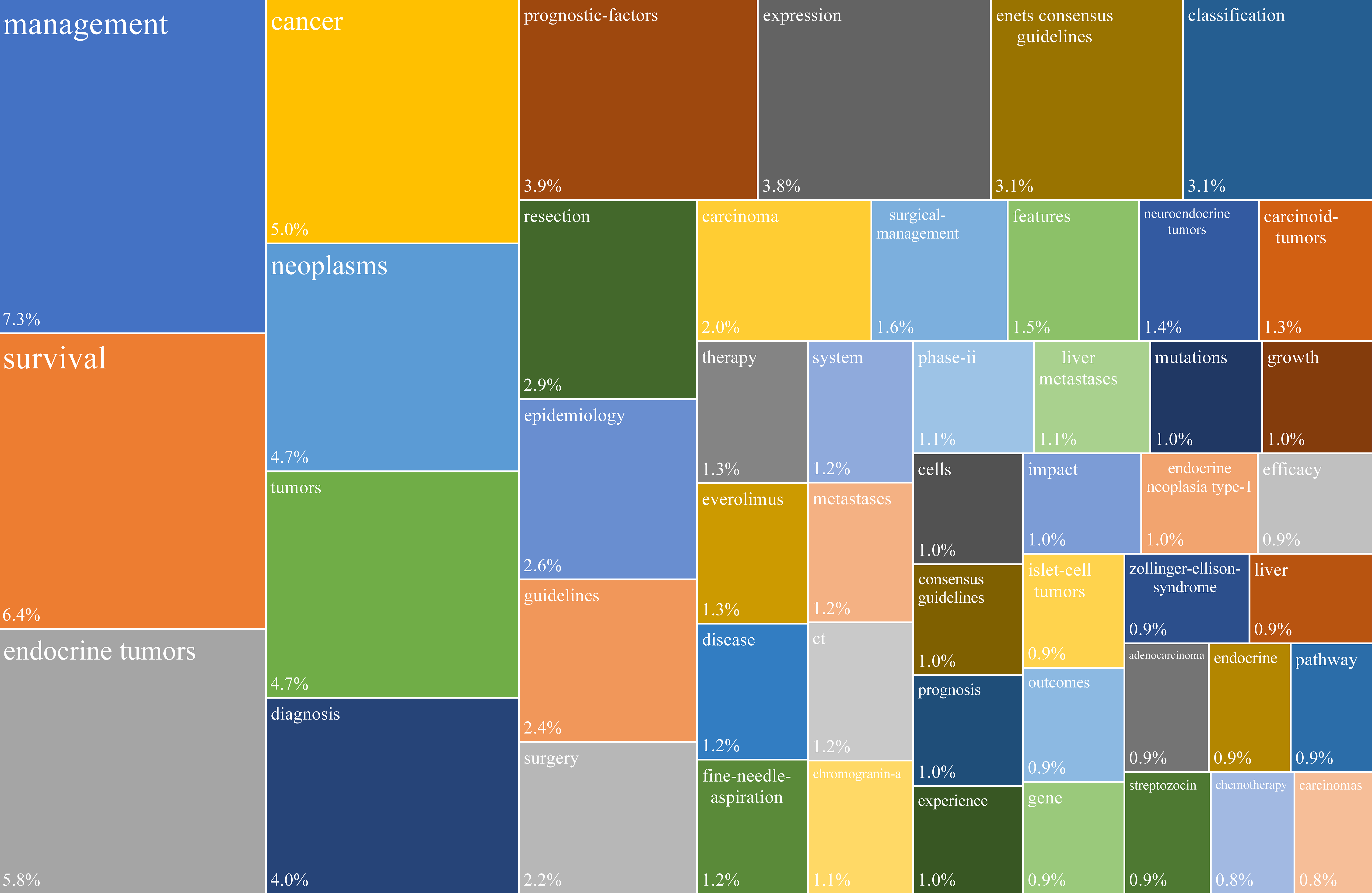
Figure 6 Treemap of the 50 most frequently occurring “Keywords Plus” terms in original articles on periampullary neuroendocrine neoplasms from 1998 to 2023.
- Citation: Zhang XY, Yu JF, Li Y, Li P. Periampullary duodenal neuroendocrine tumor in a patient with neurofibromatosis-1: A case report. World J Clin Oncol 2024; 15(9): 1222-1231
- URL: https://www.wjgnet.com/2218-4333/full/v15/i9/1222.htm
- DOI: https://dx.doi.org/10.5306/wjco.v15.i9.1222


