Copyright
©The Author(s) 2024.
World J Clin Oncol. Jul 24, 2024; 15(7): 945-952
Published online Jul 24, 2024. doi: 10.5306/wjco.v15.i7.945
Published online Jul 24, 2024. doi: 10.5306/wjco.v15.i7.945
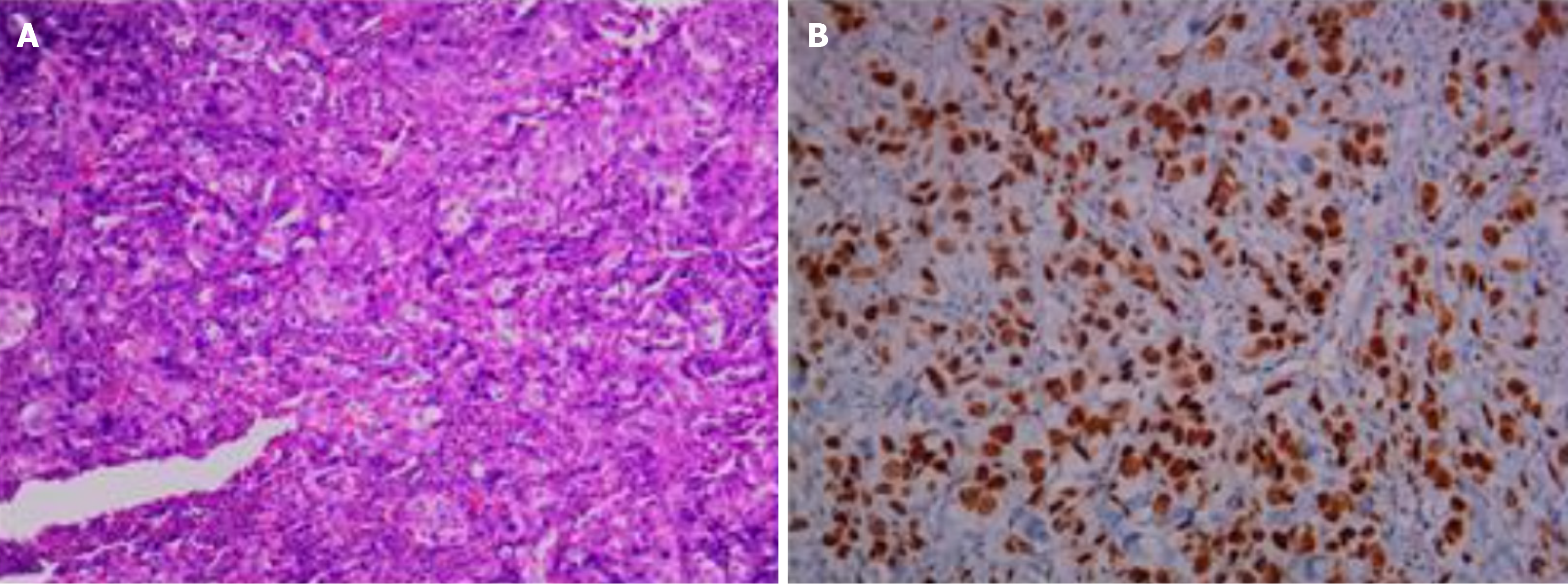
Figure 1 Histological and immunohistochemical features.
A: Histology of lung biopsy; B: Expression pattern of thyroid transcription factor 1.
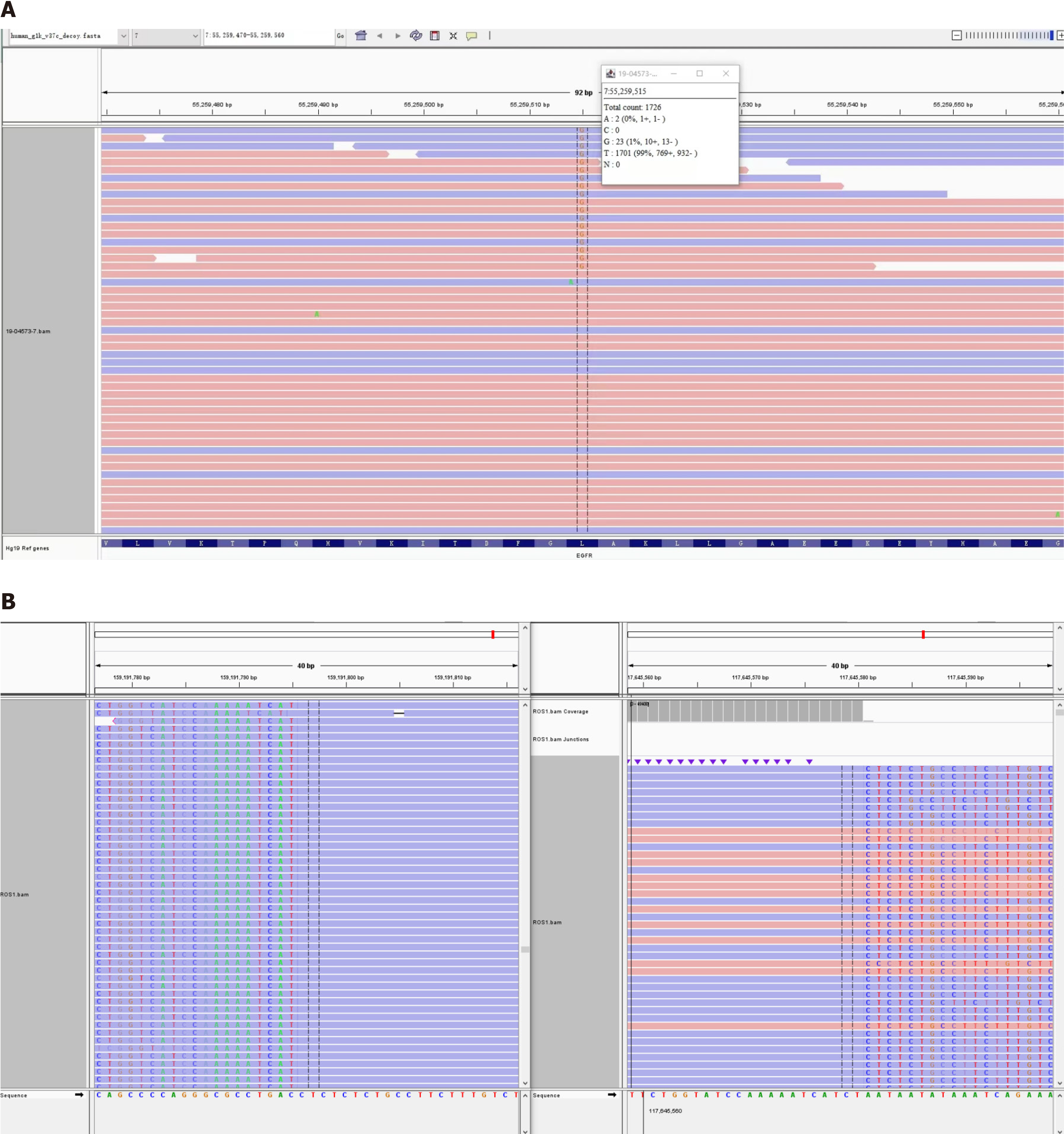
Figure 2 Detection of epidermal growth factor receptor mutation and c-ros oncogene 1 rearrangement using high sensitivity next-generation sequencing.
A: Epidermal growth factor receptor L858R mutation; B: EZR-ROS1 E10R34 fusion.
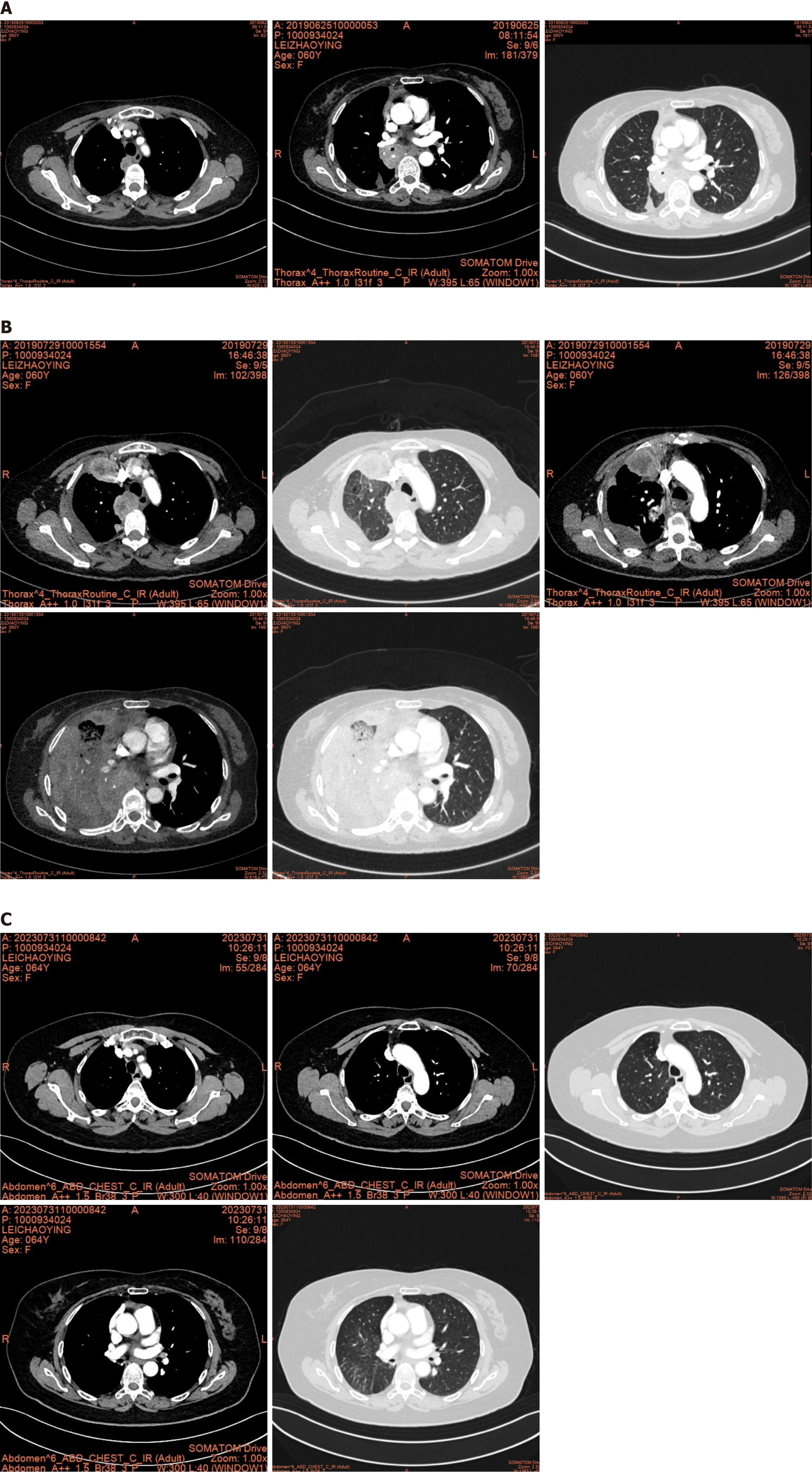
Figure 3 Serial computed tomography images.
A: Computed tomography (CT) after postoperative recurrence; B: CT after 1 month of gefitinib therapy. Progressive disease of the lung was detected; C: CT after 53 months of crizotinib therapy. Stable disease was detected.
- Citation: Peng GQ, Song HC, Chen WY. Concomitant epidermal growth factor receptor mutation/c-ros oncogene 1 rearrangement in non-small cell lung cancer: A case report. World J Clin Oncol 2024; 15(7): 945-952
- URL: https://www.wjgnet.com/2218-4333/full/v15/i7/945.htm
- DOI: https://dx.doi.org/10.5306/wjco.v15.i7.945


