Copyright
©The Author(s) 2024.
World J Clin Oncol. Jul 24, 2024; 15(7): 936-944
Published online Jul 24, 2024. doi: 10.5306/wjco.v15.i7.936
Published online Jul 24, 2024. doi: 10.5306/wjco.v15.i7.936
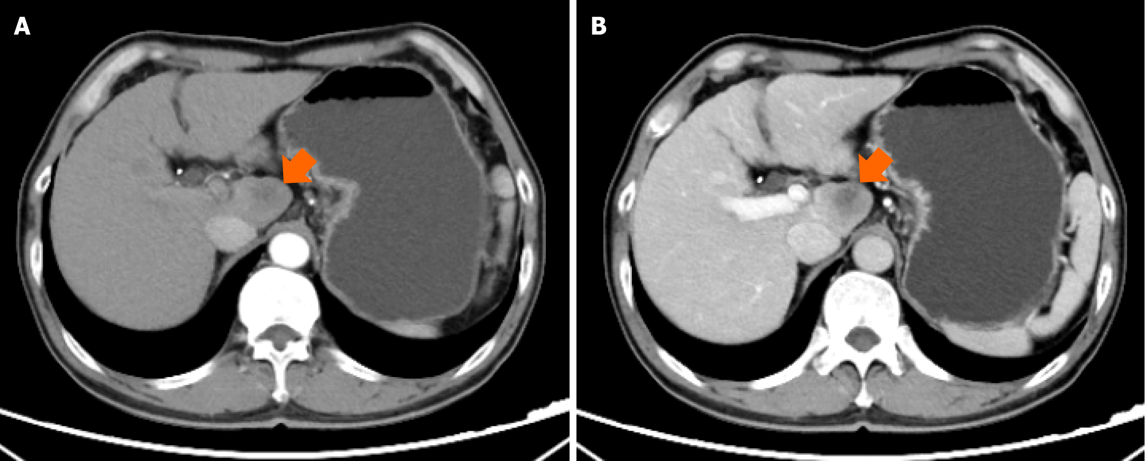
Figure 1 Computed tomography imaging of liver lesion.
A: The arterial phase of computed tomography (CT) showed no obvious enhancement (orange arrow); B: The venous phase of CT showed slightly heterogeneous enhancement (orange arrow).
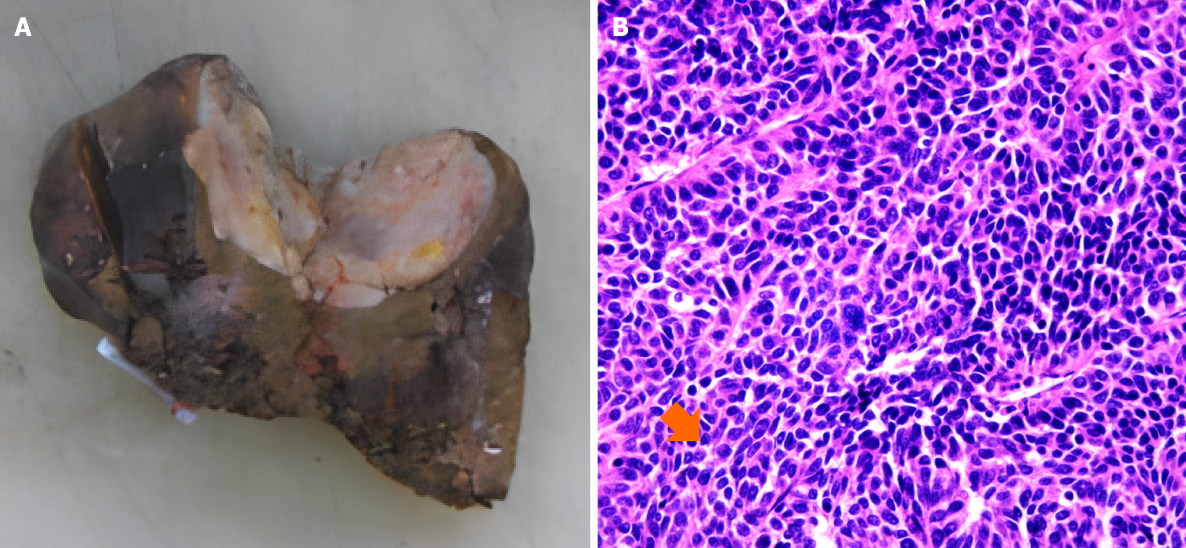
Figure 2 The pathology of liver lesion.
A: The lesion owned soft texture, partially necrosis, clear boundary, and invasion of liver capsule; B: Hematoxylin and eosin staining indicated atypical cell arranged in nests (orange arrow), and no obvious keratin pearl was observed.
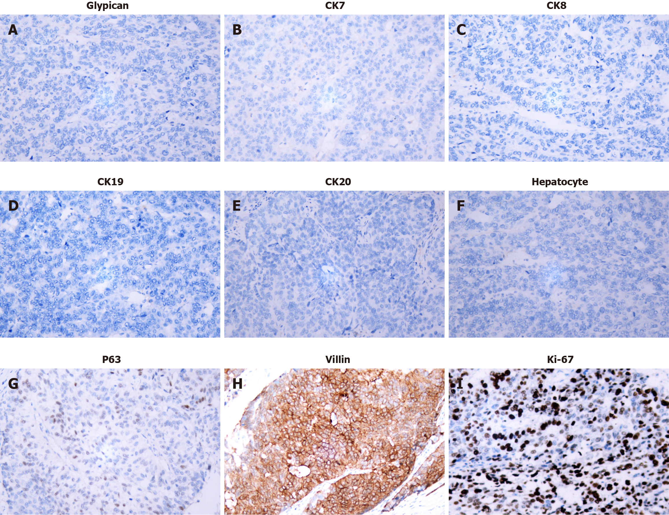
Figure 3 Immunohistochemistry of liver lesion.
The immunohistochemical examination was positive for villin, p63, and negative for glypican, hepatocyte, CK7, CK8, CK19, and CK20, the Ki-67 index was about 60% (× 200). A: Glypican; B: CK7; C: CK8; D: CK19; E: CK20; F: Hepatocyte; G: P63; H: Villin; I: Ki-67.
- Citation: Ma QJ, Wang FH, Yang NN, Wei HL, Liu F. Rare primary squamous cell carcinoma of the intrahepatic bile duct: A case report and review of literature. World J Clin Oncol 2024; 15(7): 936-944
- URL: https://www.wjgnet.com/2218-4333/full/v15/i7/936.htm
- DOI: https://dx.doi.org/10.5306/wjco.v15.i7.936


