Copyright
©The Author(s) 2024.
World J Clin Oncol. Feb 24, 2024; 15(2): 178-194
Published online Feb 24, 2024. doi: 10.5306/wjco.v15.i2.178
Published online Feb 24, 2024. doi: 10.5306/wjco.v15.i2.178
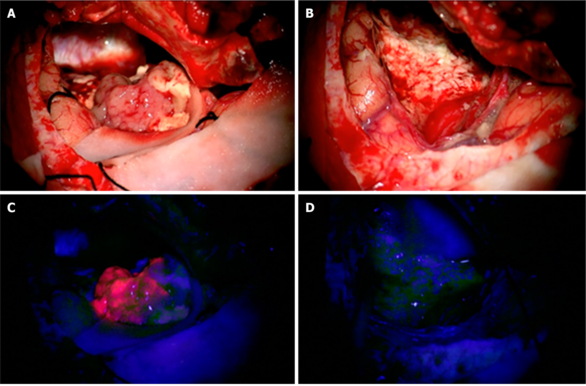
Figure 1 A 61-year-old patient with a left temporal glioblastoma.
A and B: The upper row shows the operative field in white light prior to (A) and after (B) tumor resection. C and D: The lower row shows the corresponding fluorescence images. Citation: Sales AHA, Beck J, Schnell O, Fung C, Meyer B, Gempt J. Surgical Treatment of Glioblastoma: State-of-the-Art and Future Trends. J Clin Med 2022; 11: 5354. Copyright ©2022 The Authors. Published by MDPI (Basel, Switzerland)[19] (Supplementary material).
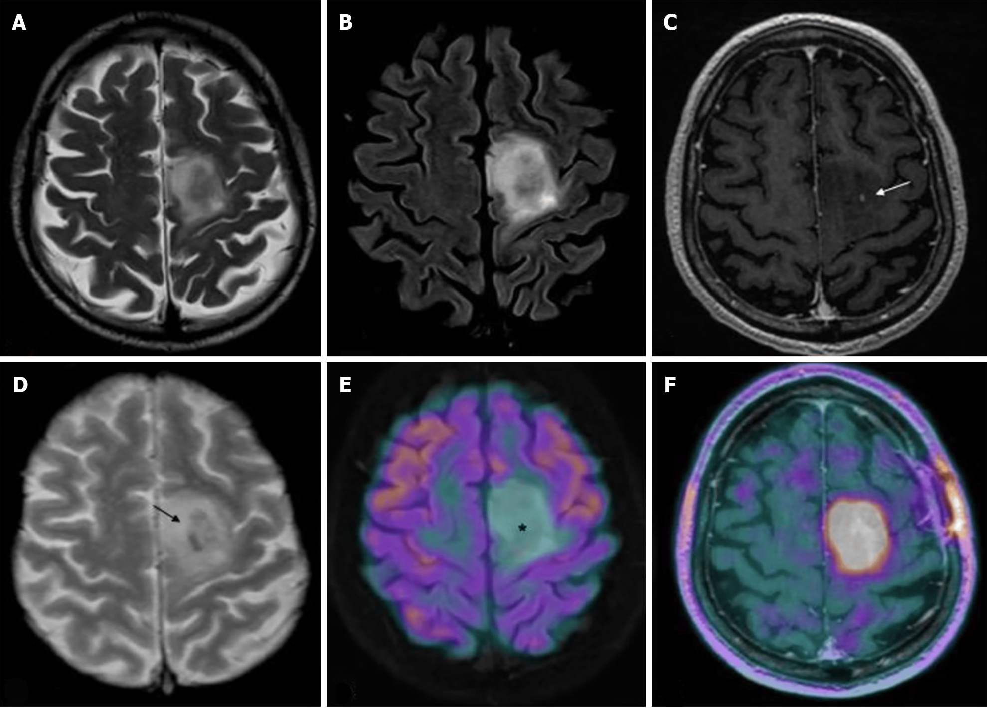
Figure 2 Magnetic resonance imaging and positron emission tomography characteristics of a diffuse isocitrate dehydrogenase mutated 1p/19q codeleted glioma (oligodendroglioma grade 2).
A-F: T2w fast spin echo (A), T2w FLAIR (B), T1w fast spin echo contrast-enhanced (C), T2*-weighted gradient echo (D), 18F-fluorodeoxyglucose (FDG)-positron emission tomography (PET)/magnetic resonance imaging (MRI) (E), and dihydroxyphenylalanine (DOPA)-PET/MRI (F) images are shown; C-F: Mesial frontal mass with a low mass effect and without any significant enhancements after contrast (arrow in C indicates the only spot of blood–brain barrier leakage), and some intratumoral calcifications (arrow in D; computed tomography not shown) with no significant radiotracer uptake by FDG-PET [asterisk in E, but showing a lively uptake of the PET-DOPA radiotracer (F)]. Citation: Feraco P, Franciosi R, Picori L, Scalorbi F, Gagliardo C. Conventional MRI-Derived Biomarkers of Adult-Type Diffuse Glioma Molecular Subtypes: A Comprehensive Review. Biomedicines 2022; 10: 2490. Copyright ©2022 The Authors. Published by MDPI (Basel, Switzerland)[28] (Supplementary material).
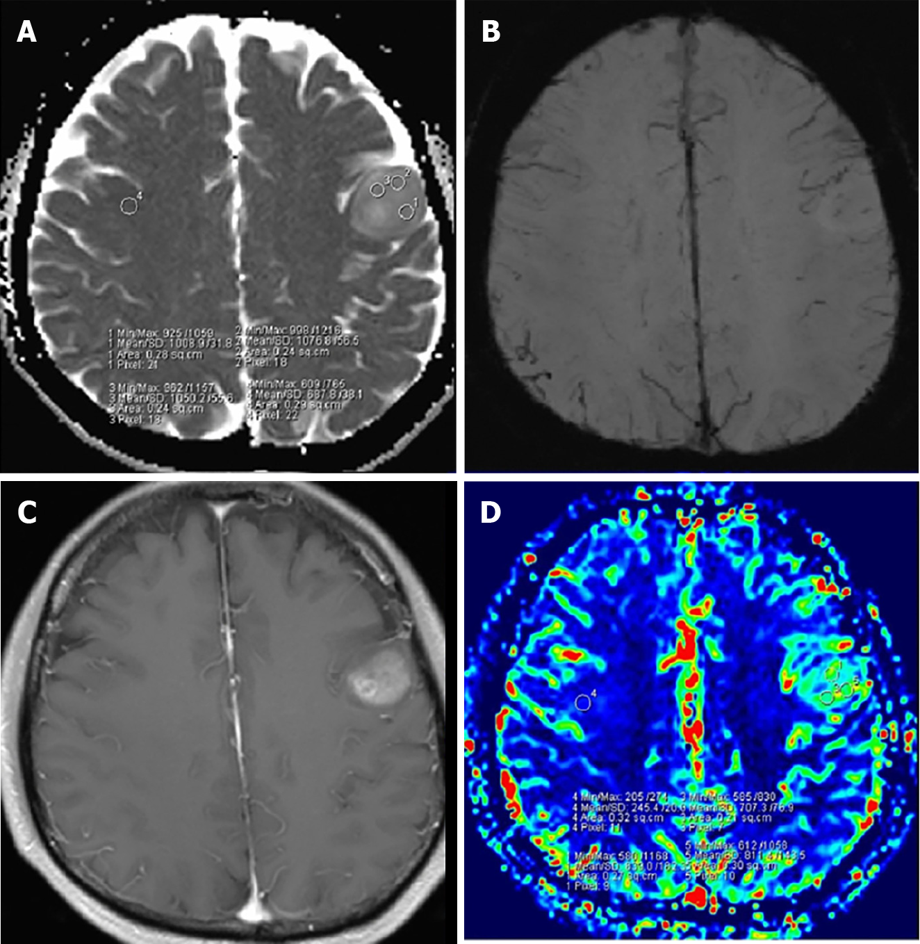
Figure 3 A 53-year-old woman with grade 3 isocitrate dehydrogenase-mutant astrocytoma.
A: Apparent diffusion coefficient (ADC) map shows an increased ADC value in the lesion (ADCmin = 1.016 × 10⁻³ mm2/s); B: Susceptibility weighted imaging shows obvious ITSS in the left parietal region; C: Post-contrast T1WI demonstrates a nonenhancing lesion; D: relative cerebral blood volume (rCBV) map shows the rCBVmax value of 1.19. Citation: Yang X, Xing Z, She D, Lin Y, Zhang H, Su Y, Cao D. Grading of IDH-mutant astrocytoma using diffusion, susceptibility and perfusion-weighted imaging. BMC Med Imaging 2022; 22: 105. Copyright ©2022 The Authors. Published by BioMed Central Ltd[31] (Supplementary material).
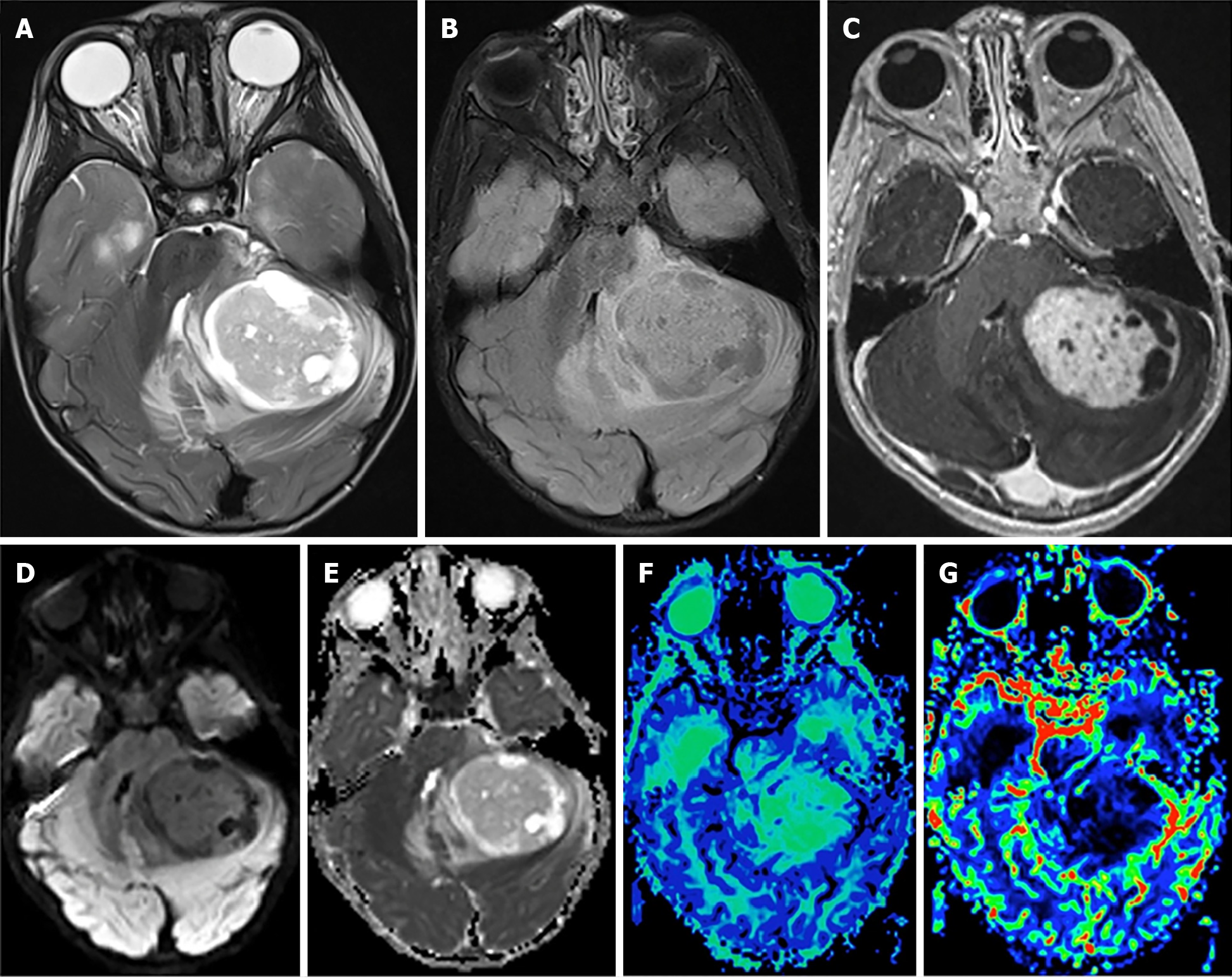
Figure 4 Pilocytic astrocytoma.
A: Axial T2-weighted scan through the cerebellum demonstrating a well-defined solid-cystic mass in the left cerebellar hemisphere that is heterogeneous onT2-weighted sequence and causes mass effect to the 4th ventricle. The solid component is minimally hyperintense compared to the gray matter; B: An axial T2 FLAIR image through the same level better demonstrates the area of T2 hyperintensity beyond the tumor margin, suggestive of edema; C: An axial post-contrast T1-weighted sequence through the same level demonstrates intense enhancement of the solid component of the tumor; D and E: The axial diffusion image and ADC map (E) through the same level show no diffusion restriction; F: The mean transit time map demonstrates increased transit time within the solid component of the tumor; G: The cerebral blood volume map shows that the tumor has very low cerebral blood volume. Citation: Bag AK, Chiang J, Patay Z. Radiohistogenomics of pediatric low-grade neuroepithelial tumors. Neuroradiology 2021; 63: 1185-1213. Copyright ©2021 The Authors. Published by Springer Nature[58] (Supplementary material).
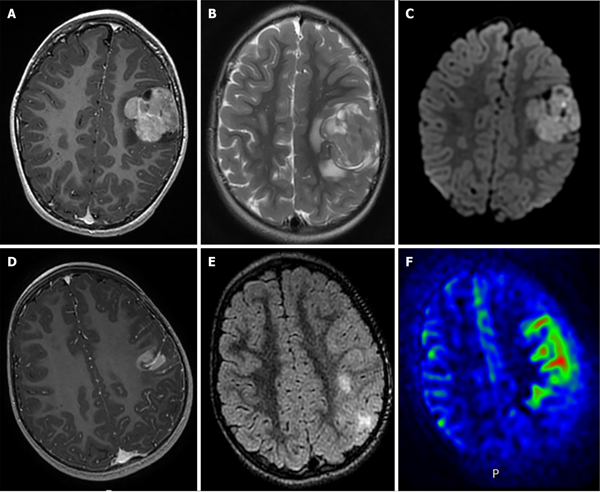
Figure 5 Radiological features of two supratentorial pediatric high-grade gliomas of the subclass MYCN.
A-C: T1-weighted images after contrast media injection (A), T2-weighted images (B), and diffusion-weighted images (C): a solid lesion with peri-lesional edema, homogeneous enhancement and hypercellularity (apparent diffusion coefficient (ADC) on diffusion weighted images is restricted in the main part of the tumor) (Case 3); D-F: T1-weighted images after contrast media injection (D), FLAIR-weighted images (E) and cerebral blood flow map using arterial spin labeling (F): a solid and infiltrative lesion with homogeneous enhancement and high cerebral blood flow (Case 1). Citation: Tauziède-Espariat A, Debily MA, Castel D, Grill J, Puget S, Roux A, Saffroy R, Pagès M, Gareton A, Chrétien F, Lechapt E, Dangouloff-Ros V, Boddaert N, Varlet P. The pediatric supratentorial MYCN-amplified high-grade gliomas methylation class presents the same radiological, histopathological and molecular features as their pontine counterparts. Acta Neuropathol Commun 2020; 8: 104. Copyright ©2020 The Authors. Published by BioMed Central Ltd[100] (Supplementary material).
- Citation: Mohamed AA, Alshaibi R, Faragalla S, Mohamed Y, Lucke-Wold B. Updates on management of gliomas in the molecular age. World J Clin Oncol 2024; 15(2): 178-194
- URL: https://www.wjgnet.com/2218-4333/full/v15/i2/178.htm
- DOI: https://dx.doi.org/10.5306/wjco.v15.i2.178


