Copyright
©The Author(s) 2024.
World J Clin Oncol. Oct 24, 2024; 15(10): 1342-1350
Published online Oct 24, 2024. doi: 10.5306/wjco.v15.i10.1342
Published online Oct 24, 2024. doi: 10.5306/wjco.v15.i10.1342
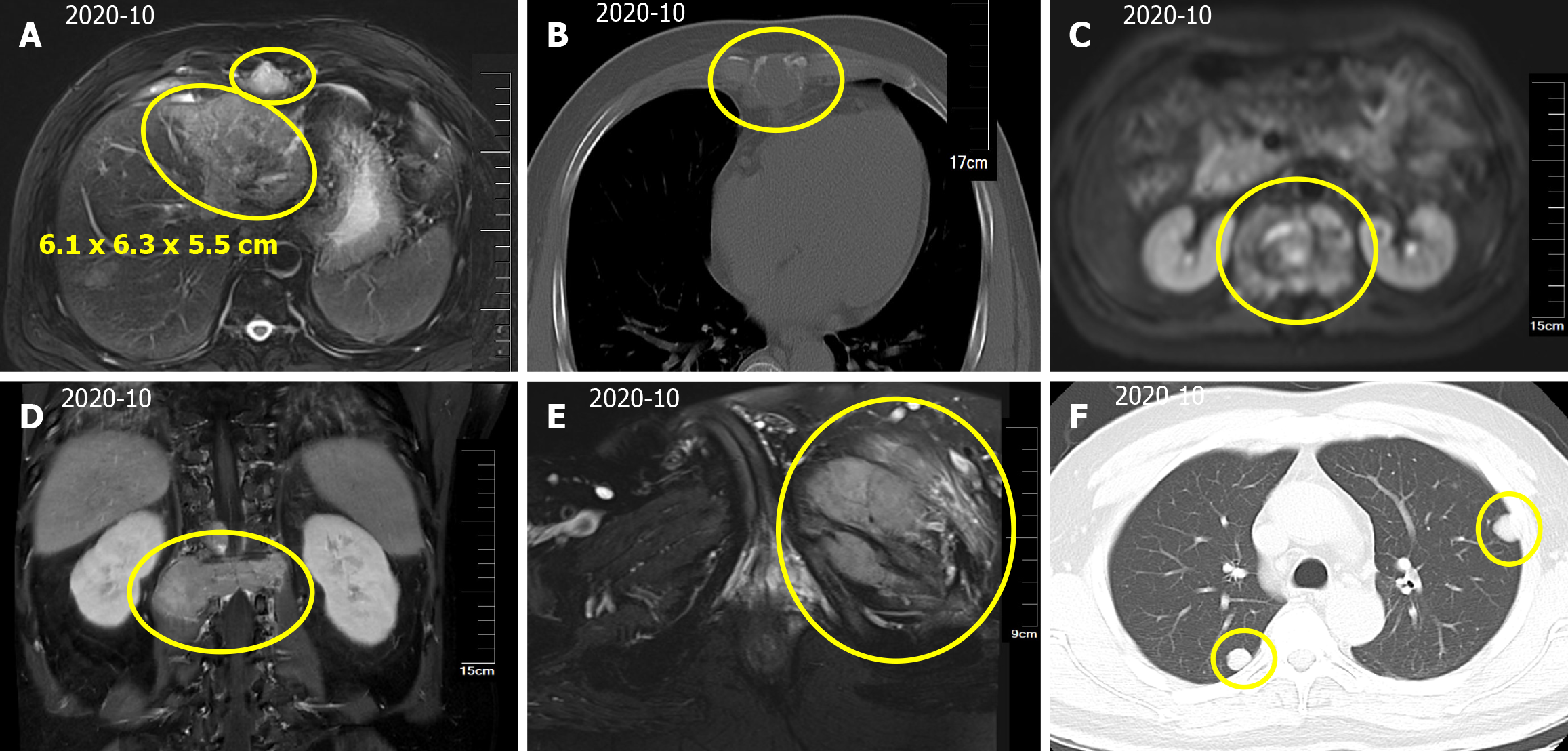
Figure 1 Hepatocellular carcinoma with intrahepatic and abdominal lymph nodes, multiple bone and lung metastases.
A: A mass in the left lobe of the liver, with a size of approximately 6.1 cm × 6.3 cm × 5.5 cm. Scattered circular abnormal signal shadows with blurred boundaries; B: Bone destruction of thoracic vertebrae and vertebral appendages; C: Paravertebral soft tissue metastases; D: L1 vertebral compression fracture, compressing spinal cord; E: Left iliac bone metastases; F: Computed tomography scan of the thorax showing multiple nodular shadows in the both lungs.
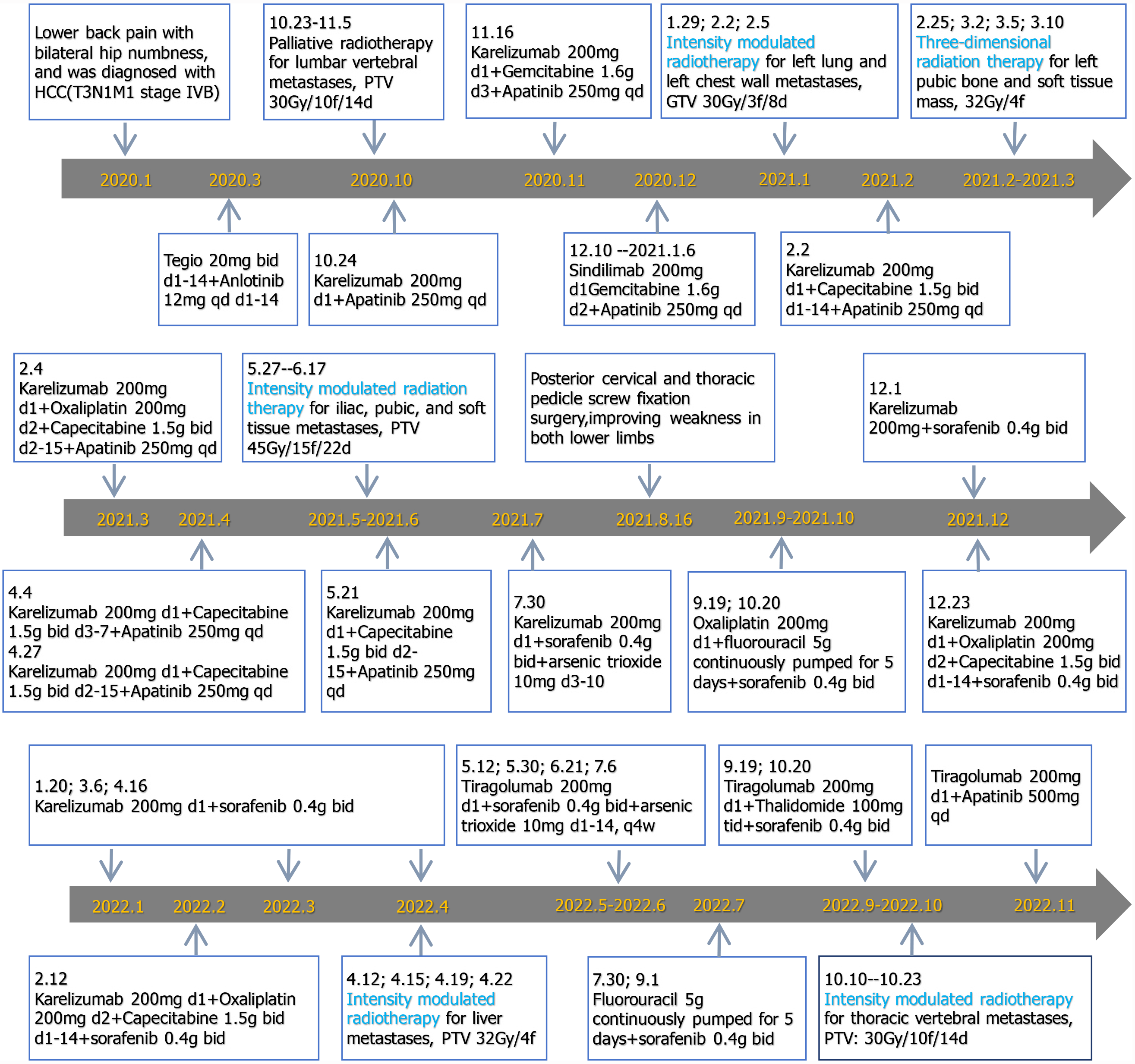
Figure 2 Detailed treatment process.
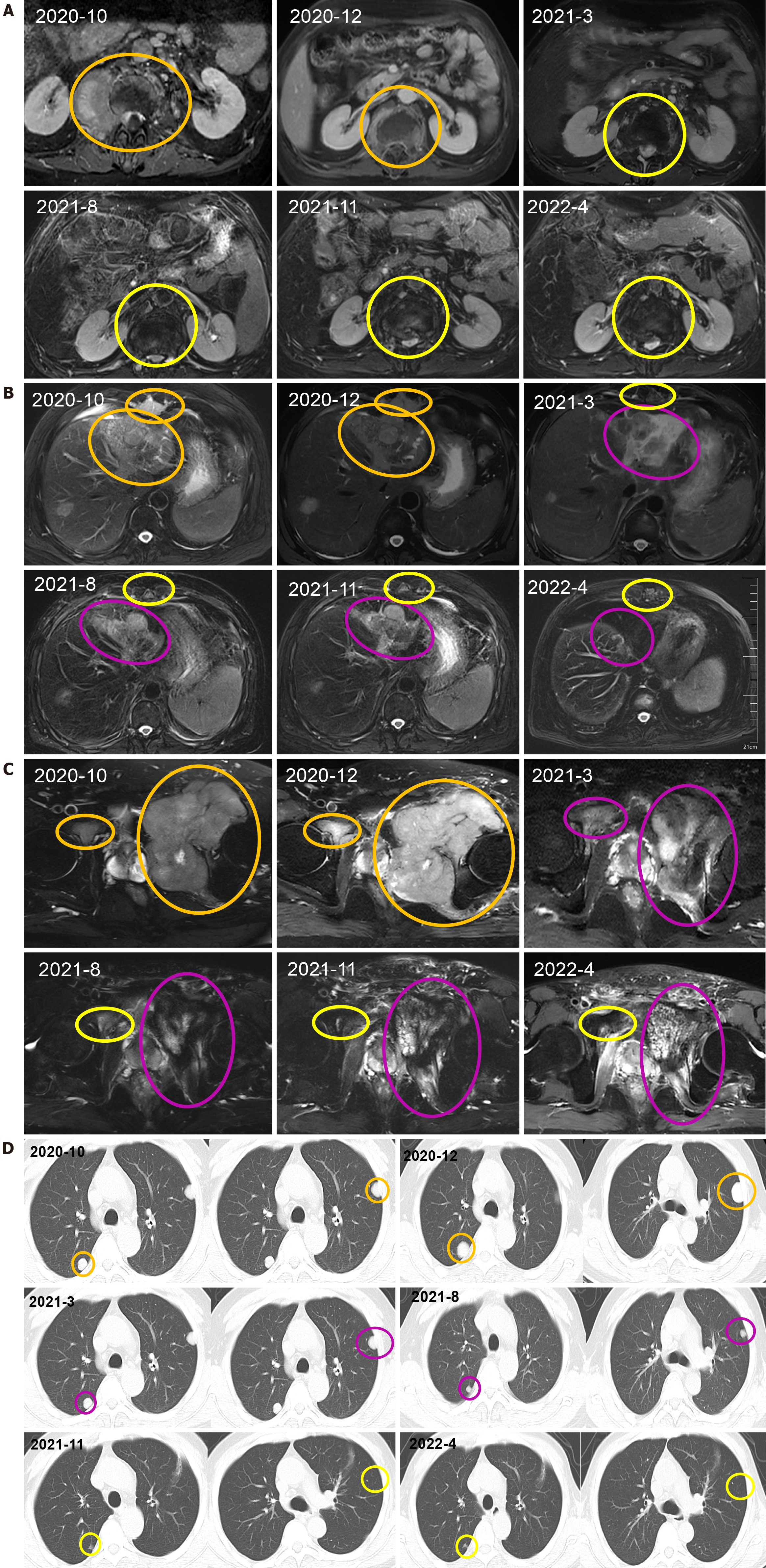
Figure 3 The primary tumor and metastatic lesions before and after therapy.
A: Vertebral body and right paravertebral mass observed via computed tomography (CT) scan; B: Liver, abdominal lymph nodes, intrahepatic lesions observed via CT scan; C: sacrum, left and right iliac bones, left iliac fossa observed via CT scan; D: CT scan of the thorax showing a nodular shadow in both lungs. All the mass was measured at the level of branching of the main pulmonary artery. The therapeutic effect was evaluated as progressive disease or stable disease are marked in orange, complete response is marked in yellow and partial response is marked in purple.
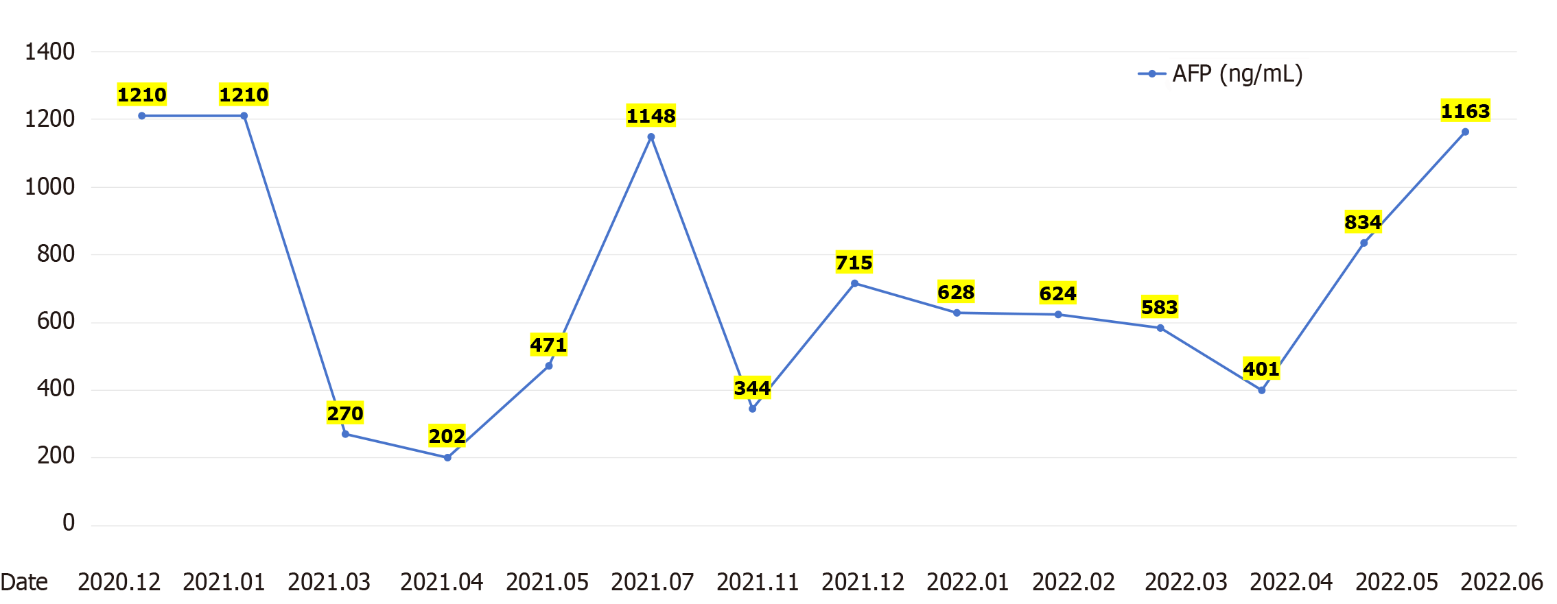
Figure 4 A-fetoprotein levels.
AFP: A-fetoprotein.
- Citation: Chen QQ, Chen CQ, Liu JK, Huang MY, Pan M, Huang H. Hypofractionated and intensity-modulated radiotherapy combined with systemic therapy in metastatic hepatocellular carcinoma: A case report. World J Clin Oncol 2024; 15(10): 1342-1350
- URL: https://www.wjgnet.com/2218-4333/full/v15/i10/1342.htm
- DOI: https://dx.doi.org/10.5306/wjco.v15.i10.1342


