Copyright
©The Author(s) 2024.
World J Clin Oncol. Oct 24, 2024; 15(10): 1315-1323
Published online Oct 24, 2024. doi: 10.5306/wjco.v15.i10.1315
Published online Oct 24, 2024. doi: 10.5306/wjco.v15.i10.1315
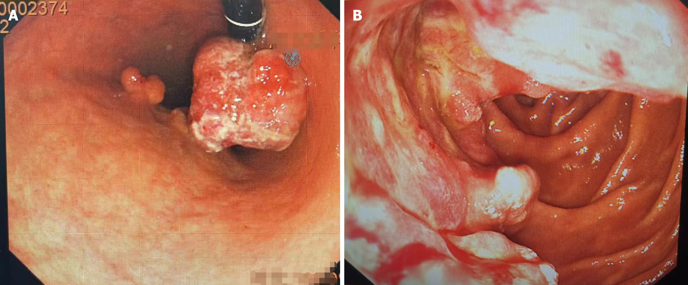
Figure 1 Patient gastroscopy.
A: Nature of multiple gastric masses; B: Duodenal carcinoma.
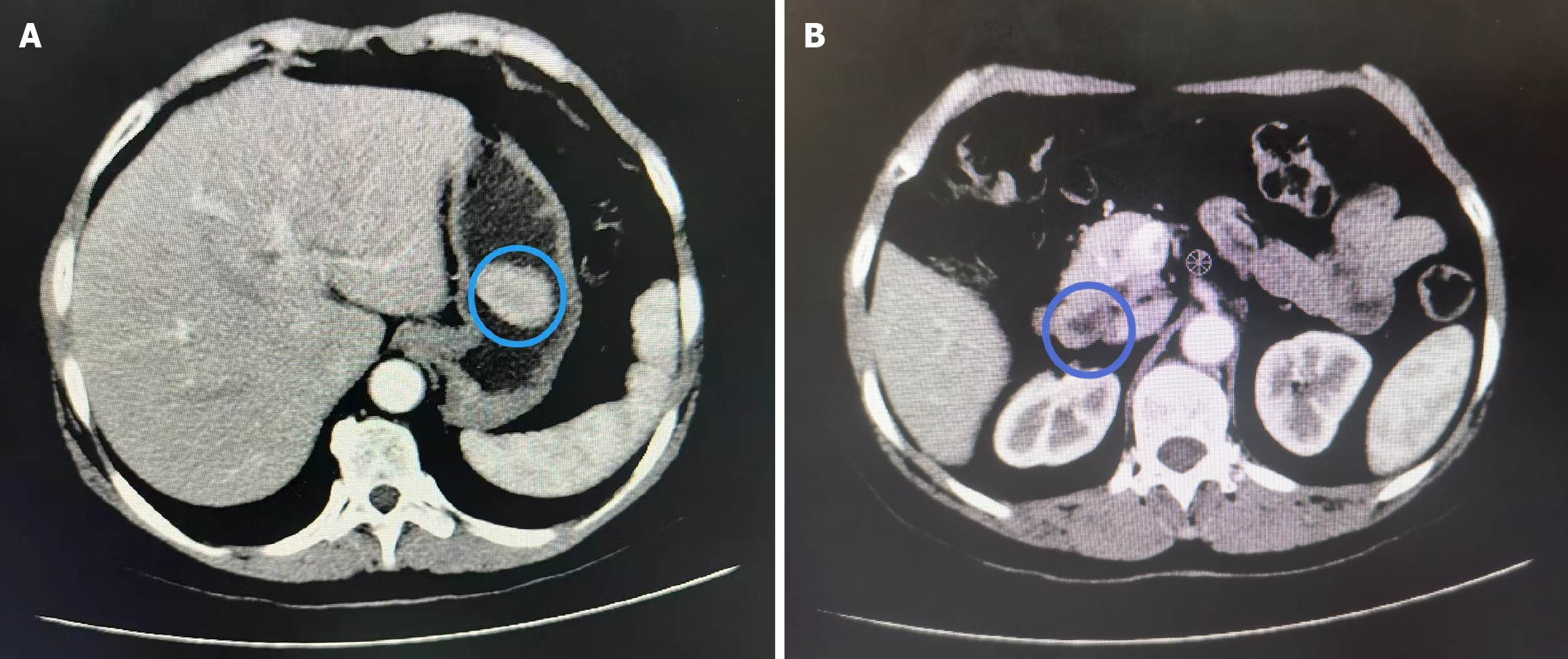
Figure 2 Patient computed tomography results.
A: Soft tissue mass in the gastric cavity; B: Thickening of the duodenal horizontal segment wall.
- Citation: Liu D, Li SC. Nursing of a patient with multiple primary cancers: A case report and review of literature. World J Clin Oncol 2024; 15(10): 1315-1323
- URL: https://www.wjgnet.com/2218-4333/full/v15/i10/1315.htm
- DOI: https://dx.doi.org/10.5306/wjco.v15.i10.1315


