Copyright
©The Author(s) 2021.
World J Clin Oncol. Jun 24, 2021; 12(6): 437-457
Published online Jun 24, 2021. doi: 10.5306/wjco.v12.i6.437
Published online Jun 24, 2021. doi: 10.5306/wjco.v12.i6.437
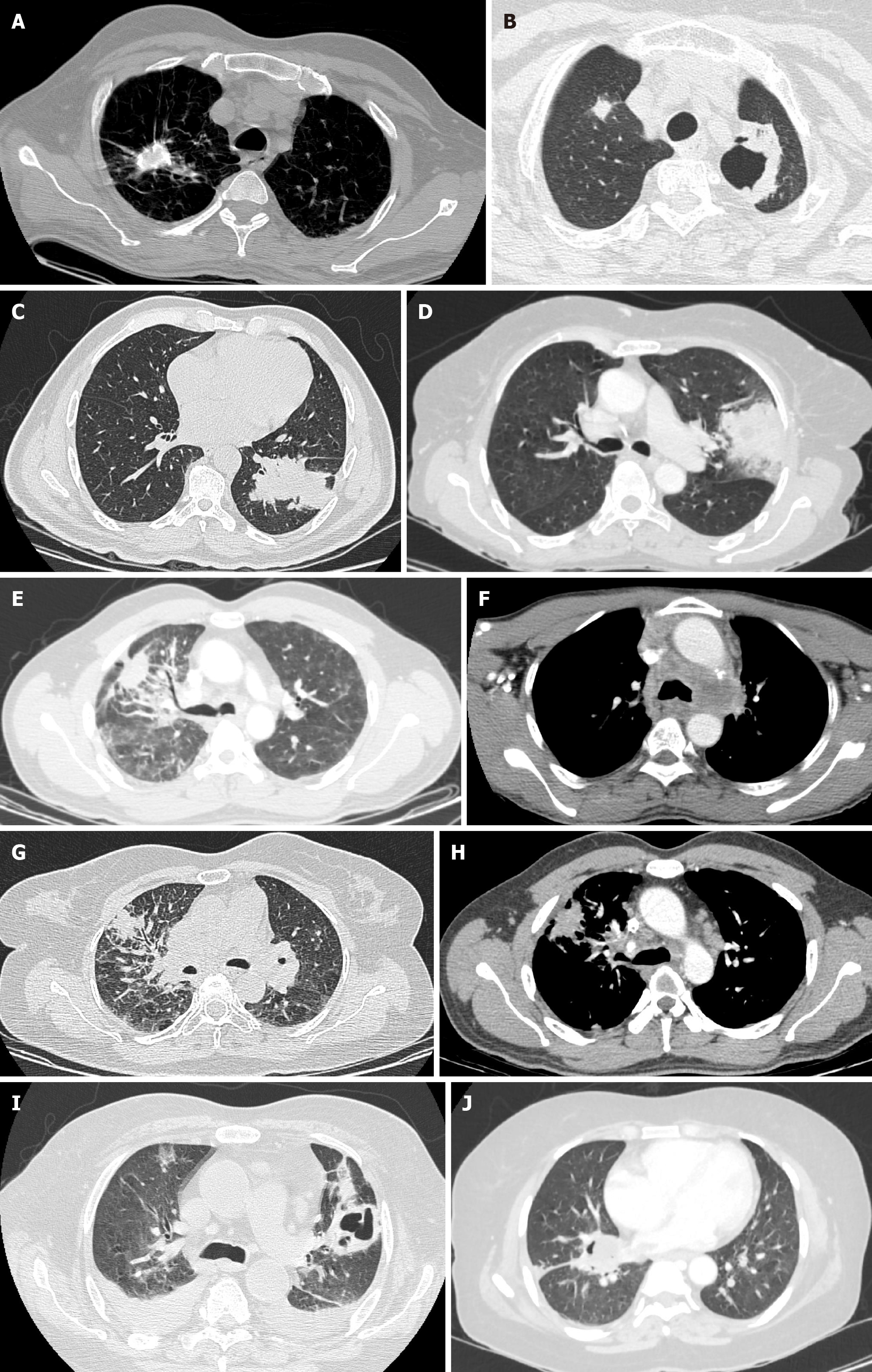
Figure 1 Axial high-resolution computed tomography images.
A: Axial high-resolution computed tomography (CT) chest image demonstrating small cell carcinoma of the right lung with dystrophic calcifications; B: Axial high-resolution CT chest image demonstrating central cavitatory squamous cell carcinoma of the left lung. Note the metastatic lesion in the right lung; C: Axial high-resolution CT chest image demonstrating large solid mass lesion with lobulated margins in a case of adenocarcinoma of the left lung; D: Axial high-resolution CT image of chest demonstrating extensive peripheral consolidation with numerous air bronchograms in a case of bronchoalveolar carcinoma of left lung; E: Axial high-resolution CT chest image demonstrating mass lesion with surrounding ground glass component representing lepidic tumor growth in a case of adenocarcinoma of the right lung; F: Axial contrast-enhanced CT image at the level of mediastinum demonstrating left hilar mass lesion with mediastinal invasion in a case of adenocarcinoma of the lung; G: Axial high-resolution CT chest image demonstrating primary bronchogenic carcinoma in the right lung with nodular and irregular interlobular septal thickening consistent with features of lymphangitis carcinomatosa; H: Axial contrast-enhanced CT chest image demonstrating sign of short burrs and spinous processes of tumor margins in a case of squamous cell carcinoma of the right lung. Note the tapered extension of the lesion to pleura and adjacent pleural retraction; I: Axial high-resolution CT chest image demonstrating peripheral cavitatory squamous cell carcinoma of the left lung. Note bilateral pleural effusions; J: Axial high-resolution CT image of chest demonstrating mass lesion invading the oblique fissure of the right lung in a case of non-small cell lung carcinoma.
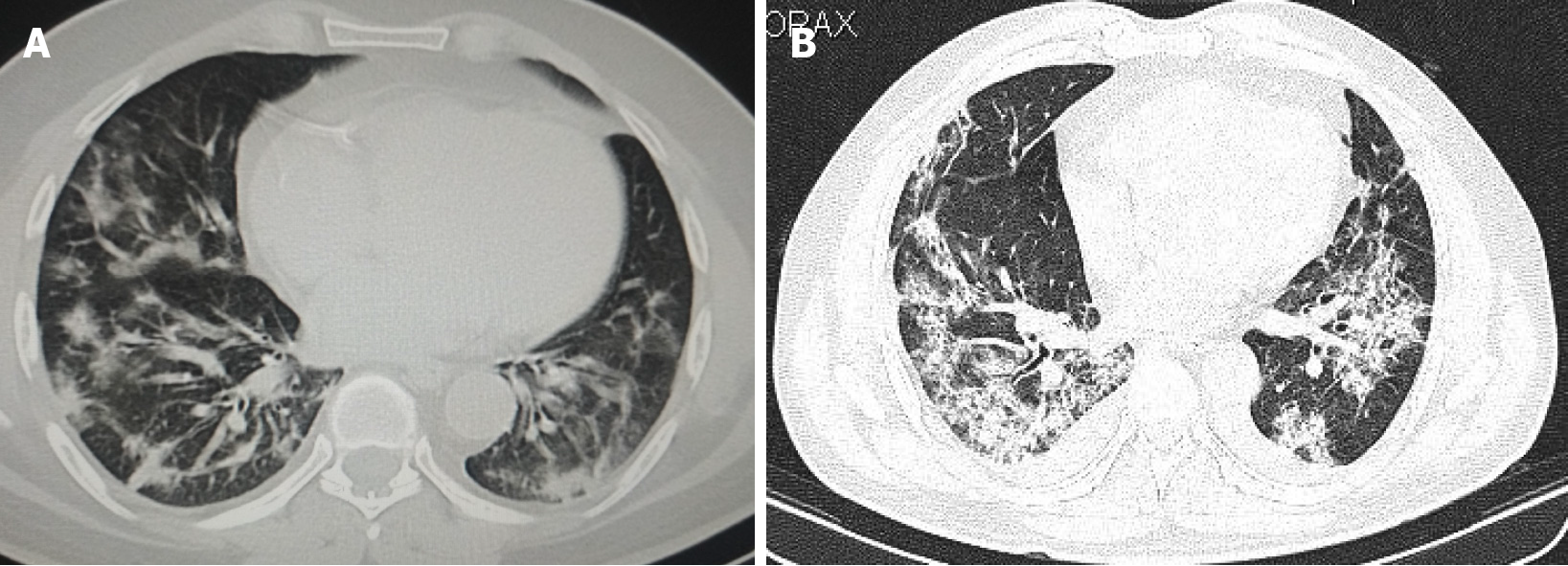
Figure 2 Axial high-resolution computed tomography images of chest.
A: Axial high-resolution computed tomography (CT) image of chest on day 5 after symptom onset demonstrating peripheral predominant consolidation pattern with areas of ground glass opacification in bilateral lower lobes in a patient with coronavirus disease 2019 pneumonia; B: Axial high-resolution CT image of chest on day 9 after symptom onset demonstrating extensive consolidation predominantly in basal segments of bilateral lower lobes in a patient with coronavirus disease 2019 related pulmonary syndrome. Note the bilateral pleural effusions which is an atypical finding in coronavirus disease 2019.
- Citation: Reddy R. Imaging diagnosis of bronchogenic carcinoma (the forgotten disease) during times of COVID-19 pandemic: Current and future perspectives. World J Clin Oncol 2021; 12(6): 437-457
- URL: https://www.wjgnet.com/2218-4333/full/v12/i6/437.htm
- DOI: https://dx.doi.org/10.5306/wjco.v12.i6.437


