Copyright
©The Author(s) 2021.
World J Clin Oncol. Feb 24, 2021; 12(2): 95-102
Published online Feb 24, 2021. doi: 10.5306/wjco.v12.i2.95
Published online Feb 24, 2021. doi: 10.5306/wjco.v12.i2.95
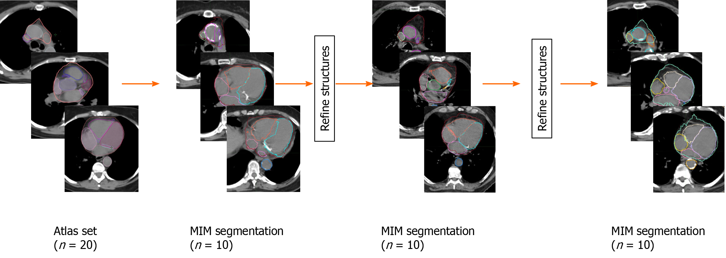
Figure 1 Project workflow of initial multi-atlas library construction, MIM autosegmentation, and subsequent edits of generated structures to add to the MIM atlas library.
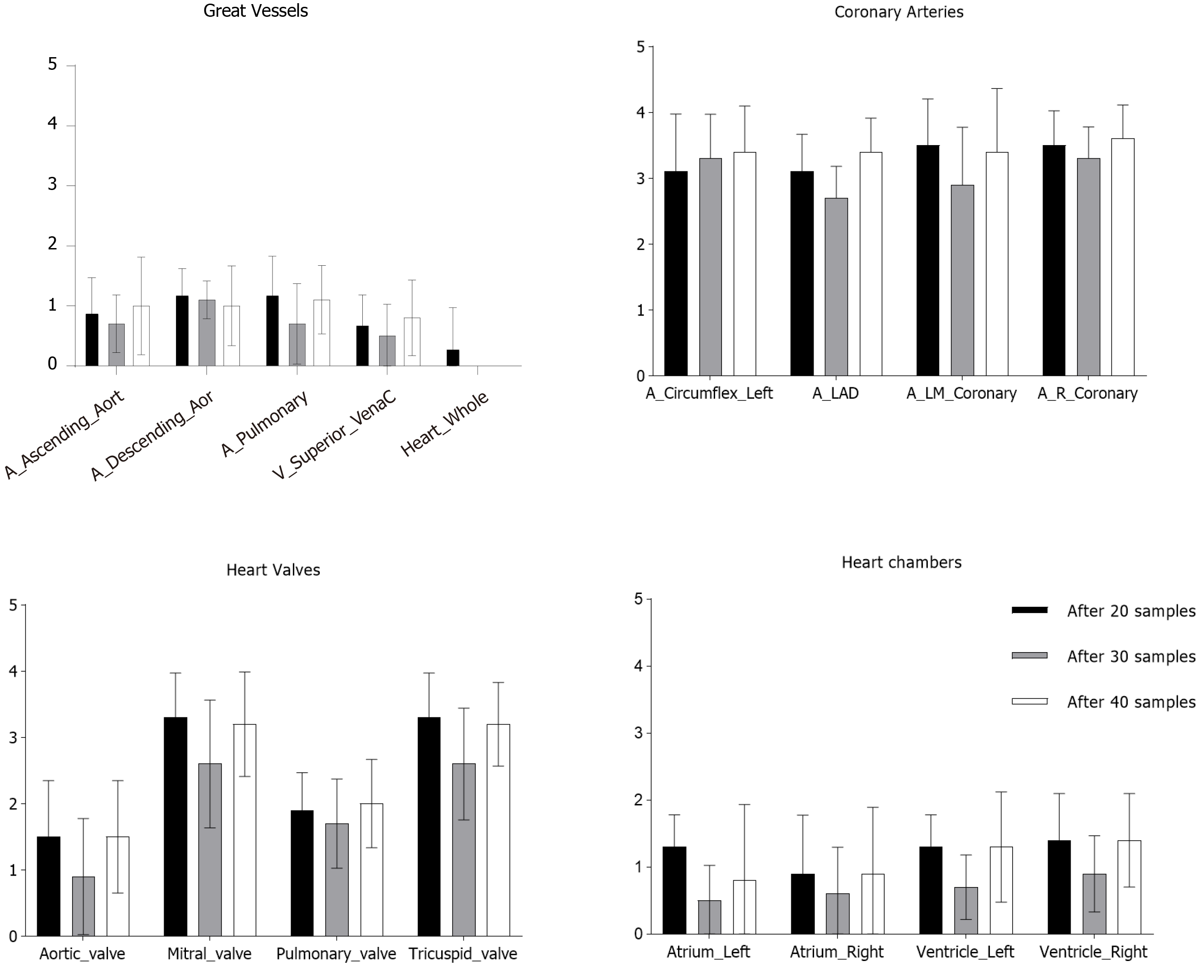
Figure 2 Physician-based evaluation of generated structures using atlas libraries comprised of 20-, 30-, and 40-patient samples (mean, standard deviation).
Scale: 0 = No changes made; 1 = Minor changes made, 2 = Moderate changes required by structure was salvageable, 3 = Structure required deletion and replacement, 4 = Structure failed autosegmentation.
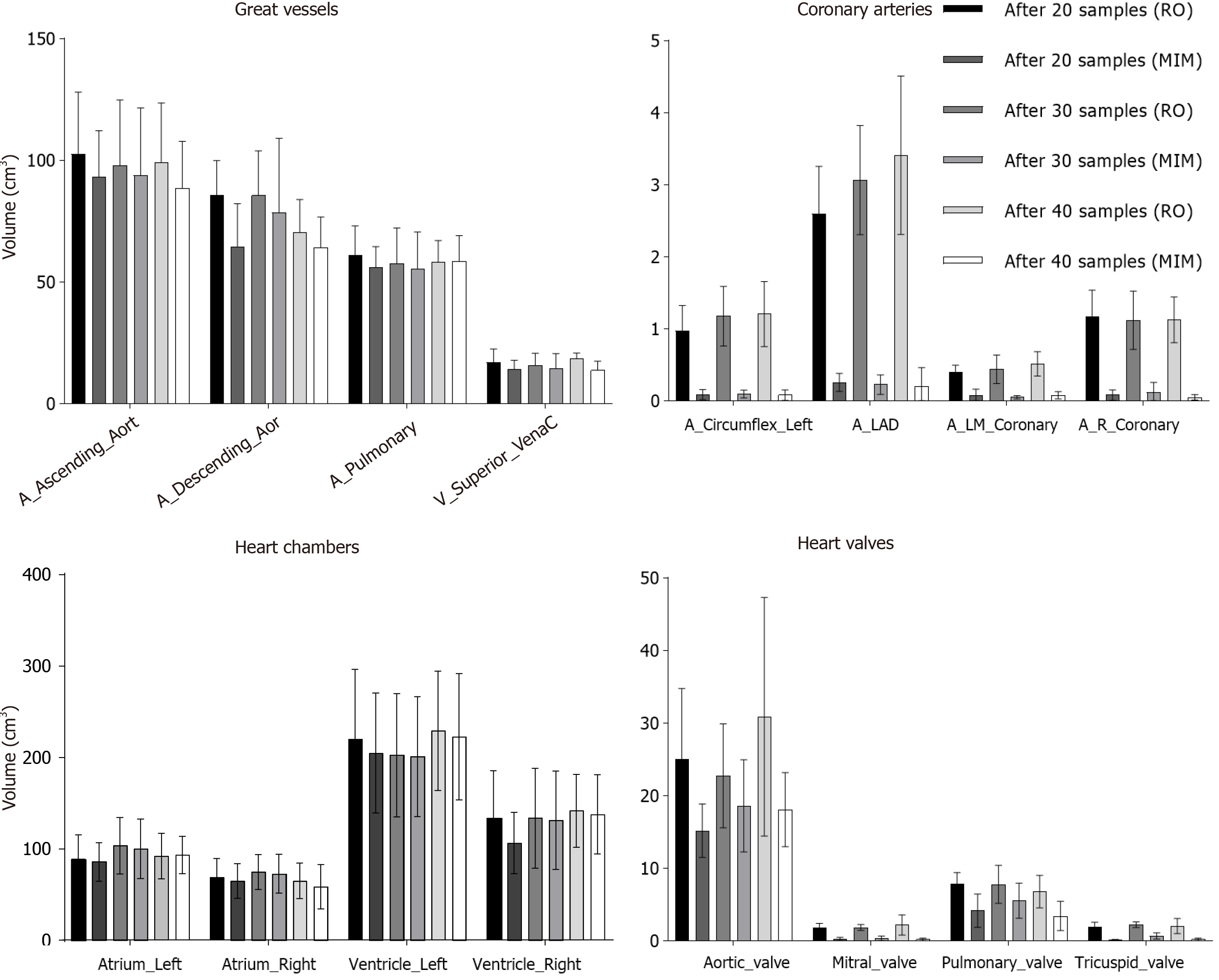
Figure 3 Analysis comparing the volumes of autosegmented structures (MIM) vs subsequent physician-edited sister (RO) (mean ± SD).
- Citation: Farrugia M, Yu H, Singh AK, Malhotra H. Autosegmentation of cardiac substructures in respiratory-gated, non-contrasted computed tomography images. World J Clin Oncol 2021; 12(2): 95-102
- URL: https://www.wjgnet.com/2218-4333/full/v12/i2/95.htm
- DOI: https://dx.doi.org/10.5306/wjco.v12.i2.95


