Copyright
©The Author(s) 2021.
World J Clin Oncol. Nov 24, 2021; 12(11): 1072-1082
Published online Nov 24, 2021. doi: 10.5306/wjco.v12.i11.1072
Published online Nov 24, 2021. doi: 10.5306/wjco.v12.i11.1072
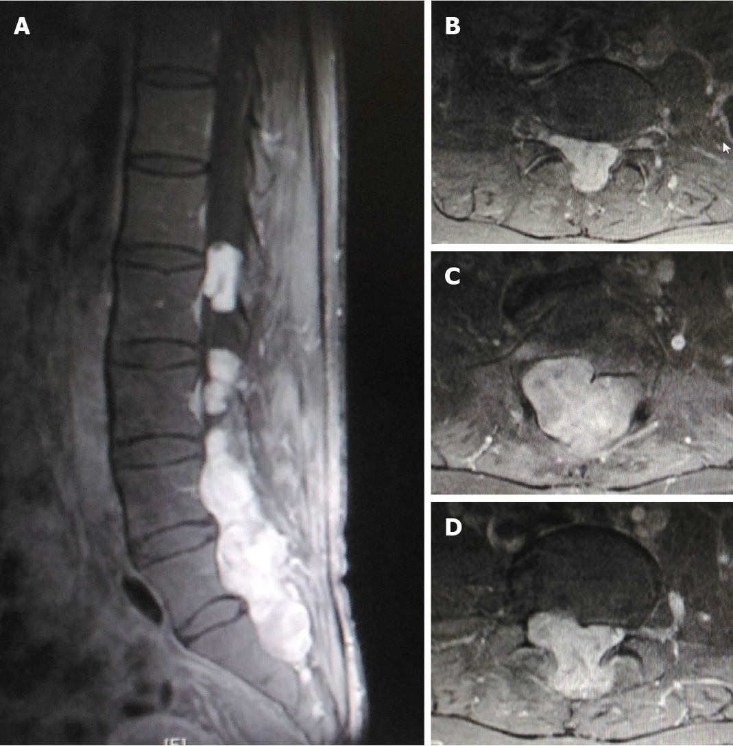
Figure 1 Gadolinium-enhanced T1 weighted magnetic resonance imaging at ultra-late recurrence of a myxopapillary ependymoma.
A: Sagittal magnetic resonance imaging (MRI) demonstrated an enhanced mass localized at L3-S1 in the intraspinal canal. A part of the mass extended into the spine axis and dorsal extraspinal tissues; B-D: Axial MRI demonstrated a slightly enhanced mass from intracanal to extraspinal regions.
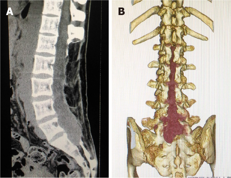
Figure 2 Computed tomography at ultra-late recurrence of a myxopapillary ependymoma.
A: A plain sagittal image demonstrated the previous laminectomy area (L1-S1); B: Three-dimensional image demonstrated a recurrent tumor (dark red) in the previous laminectomy area (L1-S1).
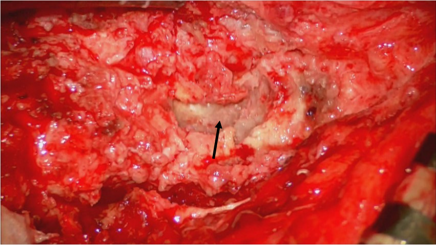
Figure 3 Intraoperative photograph showing an encapsulated tumor (arrow) surrounded by granulated tissues.
Right side: Caudal; Left side: Rostral.
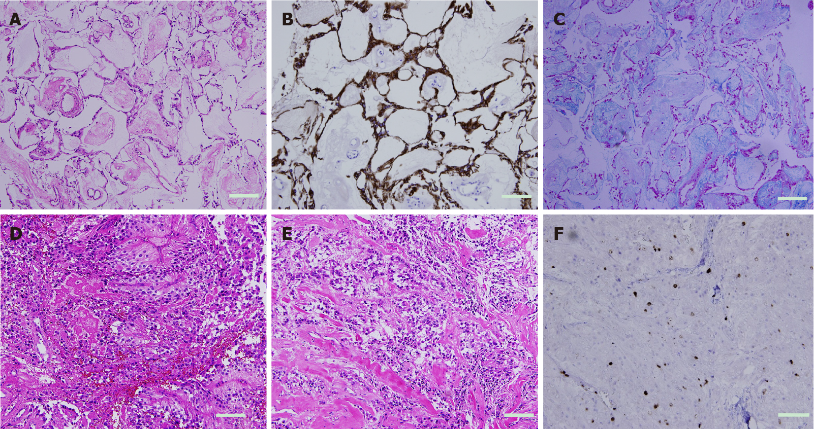
Figure 4 Microscopic appearance of the tumor.
Magnification: 200 ×; scale bar: 50 μm. A: Microphotography with hematoxylin and eosin (HE) stain showed the appearance of a typical myxopapillary ependymoma including much mucoid; B: Immunohistochemical microphotography with anti-glial fibrillary acidic protein (GFAP) antibody showed distinct expression of GFAP; C: Microphotography with Alcian blue stain showed much Alcian blue positive mucoid; D and E: Microphotography with HE stain showed anaplastic features such as hypercellularity, rapid mitotic rate, vascular proliferation, and connective tissue proliferation; F: Immunohistochemical microphotography showed a high MIB-1 labeling index (12.3%) in the area with anaplastic features.
- Citation: Kanno H, Kanetsuna Y, Shinonaga M. Anaplastic myxopapillary ependymoma: A case report and review of literature. World J Clin Oncol 2021; 12(11): 1072-1082
- URL: https://www.wjgnet.com/2218-4333/full/v12/i11/1072.htm
- DOI: https://dx.doi.org/10.5306/wjco.v12.i11.1072


