Copyright
©The Author(s) 2020.
World J Clin Oncol. Jul 24, 2020; 11(7): 504-509
Published online Jul 24, 2020. doi: 10.5306/wjco.v11.i7.504
Published online Jul 24, 2020. doi: 10.5306/wjco.v11.i7.504
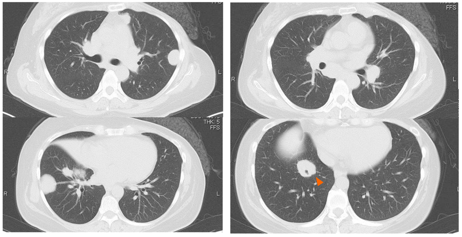
Figure 1 Chest computed tomography showing multiple bilateral masses.
Some masses bordered on the pleura and one of the masses developed around a bronchus (arrow).
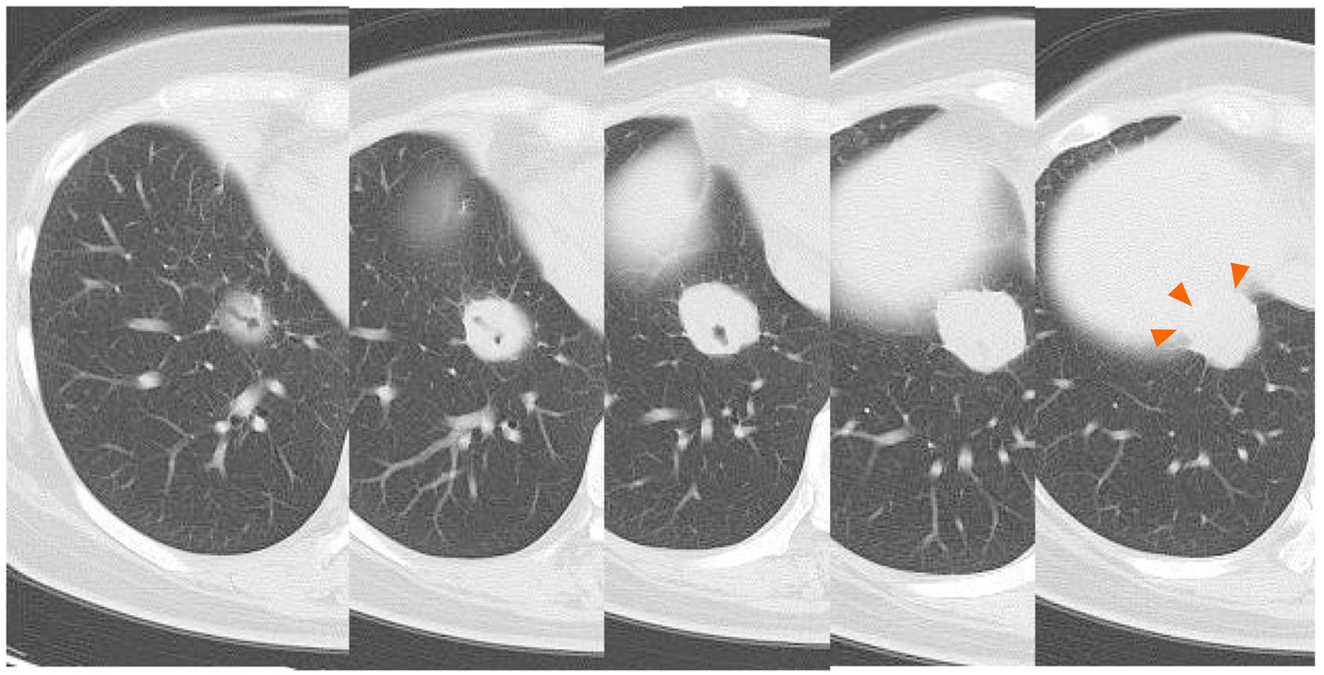
Figure 2 The mass around the bronchi is on the pleura above the diaphragm (arrows).
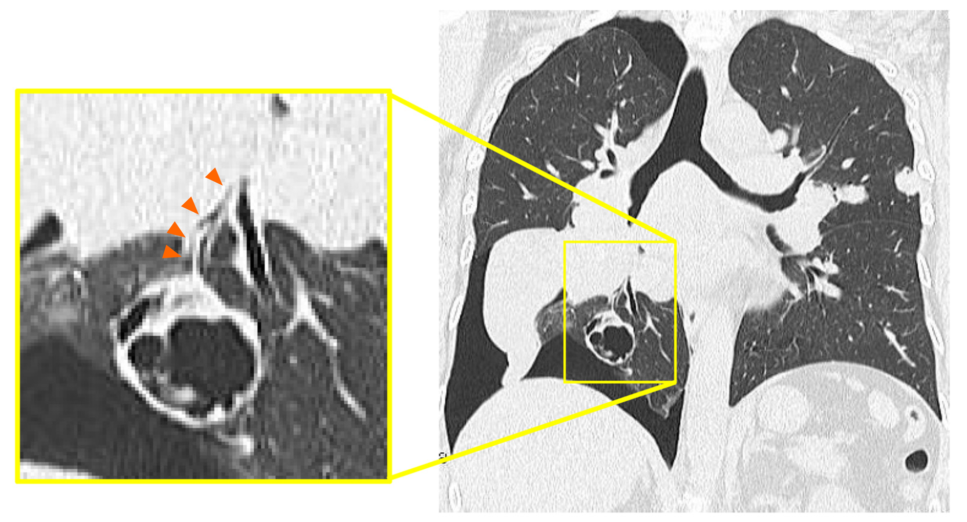
Figure 3 Coronal plane chest computed tomography showing a cavity replaced by a solid mass which was connected to the bronchi in the right lower lobe.
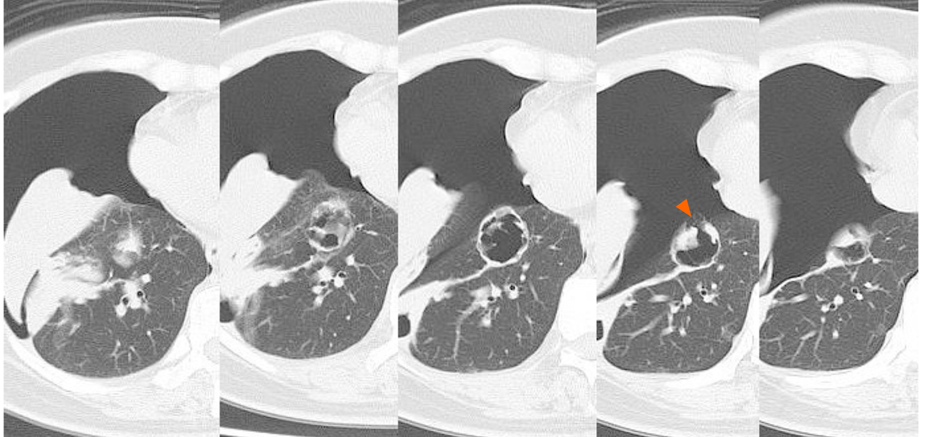
Figure 4 Chest computed tomography showing the right pneumothorax.
The cavity wall ruptured into the pleura (arrow).
- Citation: Ozaki Y, Yoshimura A, Sawaki M, Hattori M, Gondo N, Kotani H, Adachi Y, Kataoka A, Sugino K, Horisawa N, Endo Y, Nozawa K, Sakamoto S, Iwata H. Mechanisms and anatomical risk factors of pneumothorax after Bevacizumab use: A case report. World J Clin Oncol 2020; 11(7): 504-509
- URL: https://www.wjgnet.com/2218-4333/full/v11/i7/504.htm
- DOI: https://dx.doi.org/10.5306/wjco.v11.i7.504


