Copyright
©The Author(s) 2020.
World J Clin Oncol. Dec 24, 2020; 11(12): 1064-1069
Published online Dec 24, 2020. doi: 10.5306/wjco.v11.i12.1064
Published online Dec 24, 2020. doi: 10.5306/wjco.v11.i12.1064
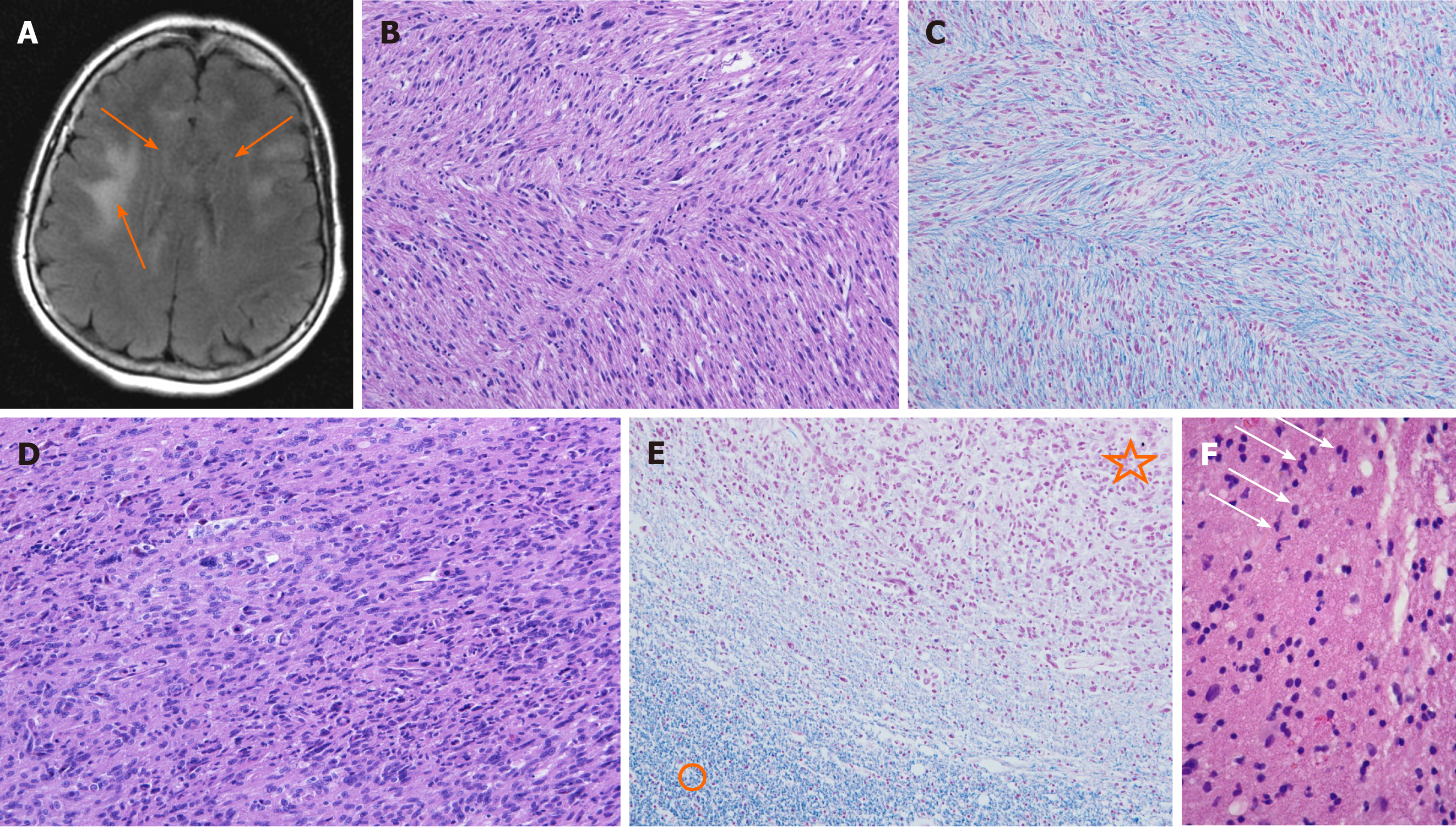
Figure 1 Magnetic resonance imaging, T2-weighted sequence, stereotactic brain biopsy, and post-mortem brain changes in the patient.
A: Magnetic resonance imaging revealed some focal areas of high intensity in the white matter of both frontal lobes (arrows) on T2-weighted sequence; B and C: Microscopic feature of cerebral gliomatosis in the genu of the corpus callosum: Hypercellular white matter with tumor cells in between the myelinated fibers [hematoxylin and eosin (H/E) staining and Klüver-Barrera staining, respectively]; D: Microscopic feature of malignant glioma in the splenium of the corpus callosum: Hypercellular tumor composed of polymorphic and some multinucleated tumor cells with brisk mitotic activity (H/E); E: There were no myelinated nerve fibers inside the tumor mass (upper part, star) compared to the adjacent white matter (lower part, circle) (Klüver-Barrera); F: Hypercellular white matter with slightly polymorphic nuclei (arrows) suspected for infiltrating neoplastic glial cells.
- Citation: Strojnik T, Kavalar R, Gornik-Kramberger K, Rupnik M, Robnik SL, Popovic M, Velnar T. Latent brain infection with Moraxella osloensis as a possible cause of cerebral gliomatosis type 2: A case report. World J Clin Oncol 2020; 11(12): 1064-1069
- URL: https://www.wjgnet.com/2218-4333/full/v11/i12/1064.htm
- DOI: https://dx.doi.org/10.5306/wjco.v11.i12.1064


