Copyright
©The Author(s) 2020.
World J Clin Oncol. Dec 24, 2020; 11(12): 1018-1028
Published online Dec 24, 2020. doi: 10.5306/wjco.v11.i12.1018
Published online Dec 24, 2020. doi: 10.5306/wjco.v11.i12.1018
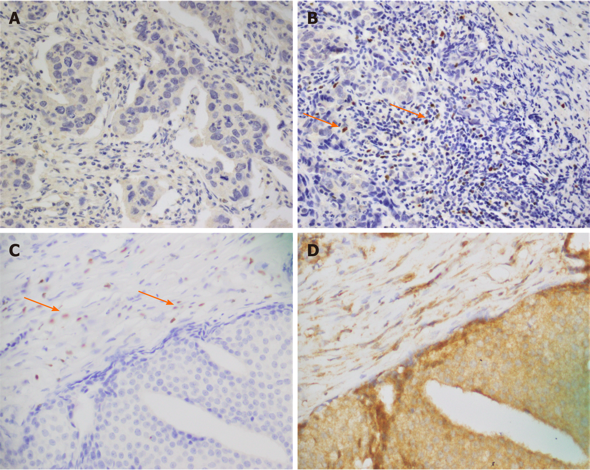
Figure 1 Formalin-fixed paraffin-embedded tumor specimens.
A and B: Forkhead box P3 (FOXP3) immunohistochemical staining; A: Invasive ductal carcinoma with FOXP3-negative expression; B: FOXP3-positive lymphocytic infiltration in invasive ductal carcinoma. The staining was nuclear; C and D: Co-expression of FOXP3 and indoleamine 2,3-dioxygenase (IDO), FOXP3 and IDO expression in breast cancer tissues (n = 100) were evaluated; C: FOXP3 positive cells infiltrated in invasive ductal carcinoma (nuclear staining). Sections from matched breast cancer patients were stained for IDO; D: Strong and diffuse IDO staining in invasive ductal tumor cells (cytoplasmic staining). Images were captured at × 40 magnification.
- Citation: Asghar K, Loya A, Rana IA, Bakar MA, Farooq A, Tahseen M, Ishaq M, Masood I, Rashid MU. Forkhead box P3 and indoleamine 2,3-dioxygenase co-expression in Pakistani triple negative breast cancer patients. World J Clin Oncol 2020; 11(12): 1018-1028
- URL: https://www.wjgnet.com/2218-4333/full/v11/i12/1018.htm
- DOI: https://dx.doi.org/10.5306/wjco.v11.i12.1018


