Copyright
©The Author(s) 2020.
World J Clin Oncol. Oct 24, 2020; 11(10): 761-808
Published online Oct 24, 2020. doi: 10.5306/wjco.v11.i10.761
Published online Oct 24, 2020. doi: 10.5306/wjco.v11.i10.761
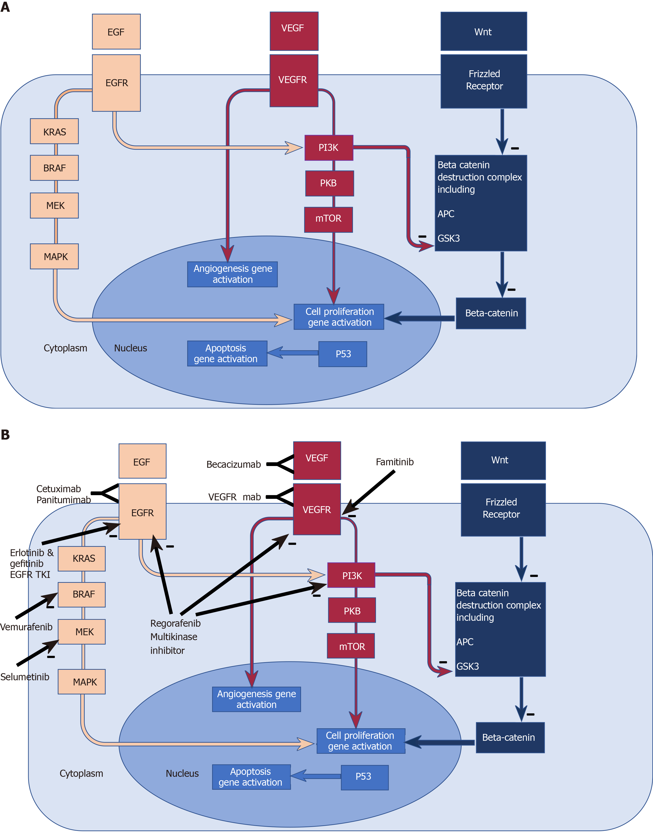
Figure 1 Biomarkers, molecular pathways, existing and emerging therapeutic targets in colorectal cancer.
A: Biomarkers and molecular pathways in colorectal cancer. Epidermal Growth Factor Receptor (EGFR) pathway: EGFR is a transmembrane receptor tyrosine kinase[340]. EGF binding to the extracellular domain results in activation of down- stream intracellular signalling pathways such as RAS-RAF-MEK-MAPK, and the PI3K PKB mTOR pathway, amongst others, which favour cell proliferation and survival[341-344]; Angiogenesis pathway: Vascular endothelial growth factors influence angiogenesis in health and disease via binding to the vascular endothelial growth factor receptor. Deregulated angiogenesis impacts on progression in solid tumours, thus providing potential anti-angiogenic therapies[345]; Wnt pathway: The Wnt genes are vast family of highly conserved genes with wide ranging roles in development, cell proliferation and migration and tumorigenesis[346]. Beta catenin accumulation in the cytoplasm and nucleus leads to cell proliferation. Excess beta catenin accumulation is prevented by its destruction by the “beta catenin destruction complex” (a multiprotein assembly containing adenomatous polyposis coli and GSK3). Wnt binding to its receptor frizzled leads to impaired function of the Beta catenin destruction complex and hence beta catenin accumulation and cell proliferation[347]. Mutations in adenomatous polyposis coli prevent the formation of the beta catenin complex, and therefore allow beta catenin accumulation and cell proliferation. PI3K inhibits the function of GSK3[112], thereby impairing the beta catenin destruction complex, hence contributing to the tumorigenic accumulation of beta catenin. B: Existing and emerging therapeutic targets in colorectal cancer pathways. B: Cetuximab and Panitumumab are monoclonal antibodies targeting the EGFR, thus blocking activation of downstream signalling pathways. Mutated and constitutively active downstream effectors (such as RAS and RAF) confer resistance to EGFR blockade. Erlotinib and gefitinib are EGFR Tyrosine kinase inhibitors and are associated with improved PFS when combined to Bevacizumab in the DREAM trial[156]. Vemurafenib is a RAF inhibitor which in combination with EGFR blockade[157] has shown marked responses in some case reports[158]. Selumetinib is a MEK kinase inhibitor showing tumour response in some patients with KRAS mutant colorectal cancers progressing on Oxaliplatin[159]. Regorafenib inhibits is a multi-kinase inhibitor[153], with OS benefit in randomised double blind control trials[154,155]. Bevacizumab is a Monoclonal antibody against VEGFA with the most prominently established role in the treatment of metastatic colorectal cancer. Famitinib is a multiple tyrosine kinase inhibitor and targets the vascular endothelial growth factor receptor tyrosine kinase. Monoclonal antibodies targeting the VEGF receptors are also under investigation[160]. EGF: Epidermal Growth Factor; EGFR: Epidermal Growth Factor Receptor; EGFR TKI: Epidermal Growth Factor Receptor tyrosine kinase inhibitor; K-ras: K-Ras protein (product of the proto-oncogene KRAS); BRAF: BRAF protein (product of the proto-oncogene BRAF; MEK: Mitogen activated protein kinase which activates MAPK (mitogen-activated protein kinase); VEGF: Vascular endothelial growth factor; VEGFR: Vascular endothelial growth factor receptor; PI3K: phosphoinositide 3-kinase; PKB: Protein kinase B; mTOR: Mammalian target of Rapamycin; Wnt: Wnt protein product of proto-oncogne Wnt; Frizzled Receptor: Receptor for Wnt; APC: Protein product of the tumour suppressor gene APC (Adenomatous polyposis coli); GSK3: Glycogen synthase kinase 3.
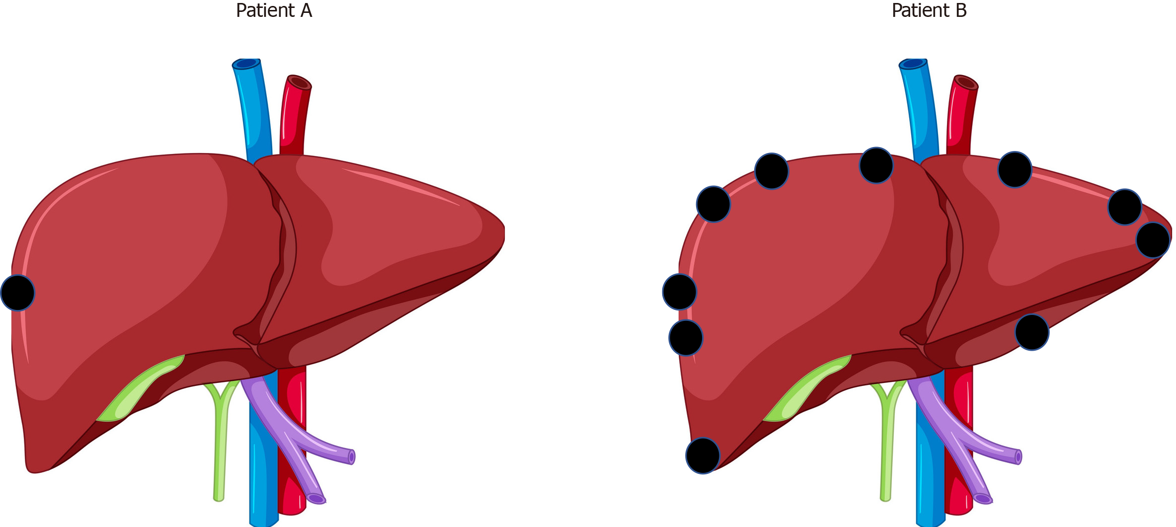
Figure 2 A hypothetical example is shown.
A: Patient A (with a single superficial colorectal liver metastases); B: Patient B (with 10 superficial colorectal liver metastases).
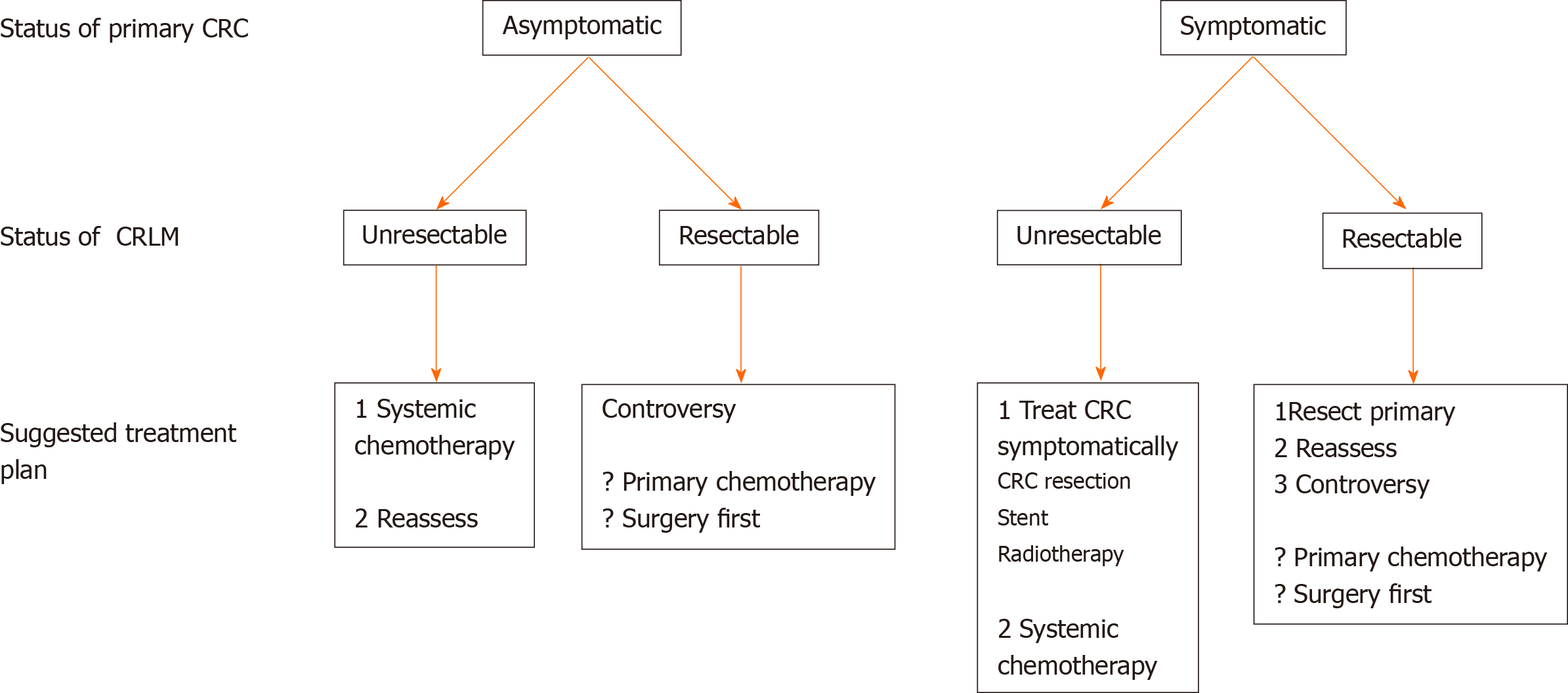
Figure 3 Management options in synchronous colorectal cancer and colorectal liver metastases, according to presentation criteria.
CRC: Colorectal cancer; CRLM: Colorectal liver metastases.
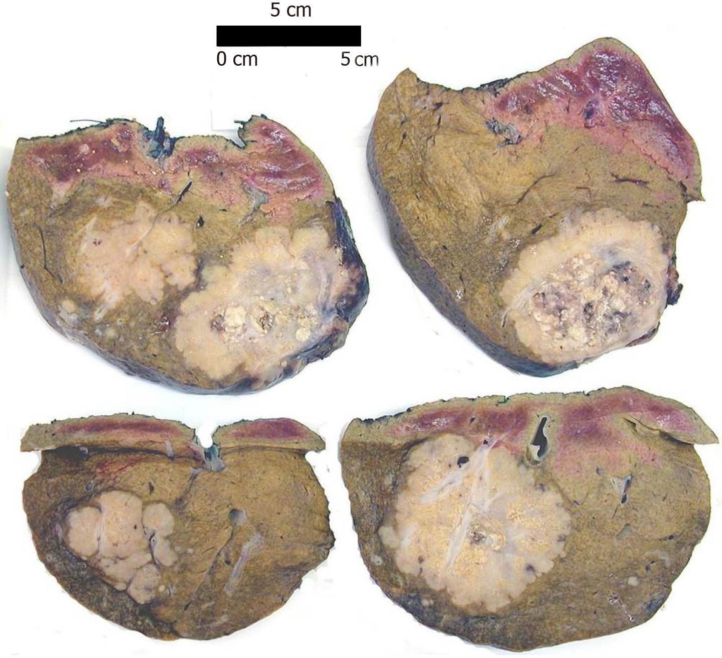
Figure 4 Multiple colorectal metastases in a formalin-fixed liver resection specimen showing a pale cut surface with typical lobulated border.
Non-capsular surfaces inked for margin identification (scale bar: 5 cm).
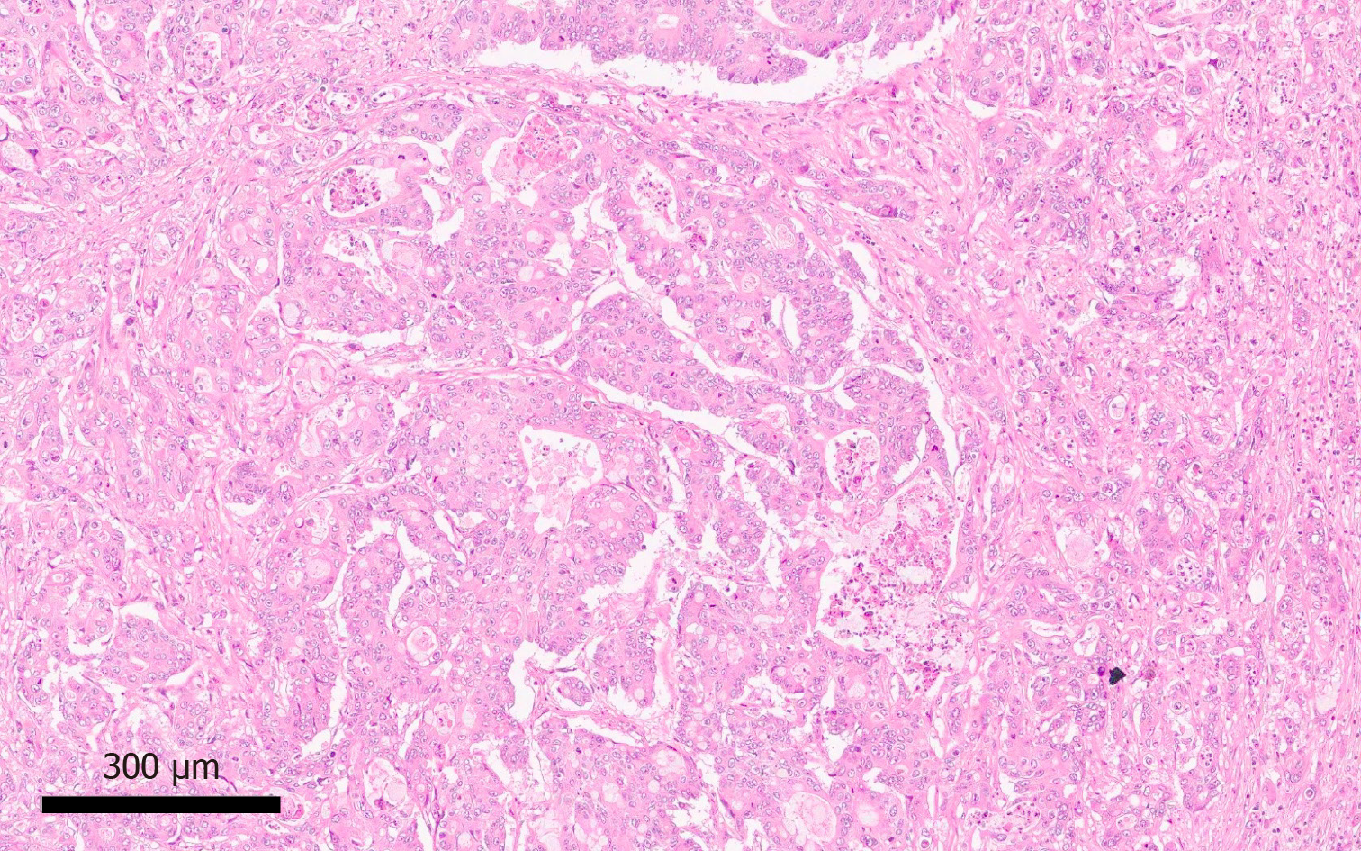
Figure 5 Colorectal metastasis with typical cribriform glandular architecture and central comedonecrosis (Hematoxylin-eosin staining, × 10 magnification).
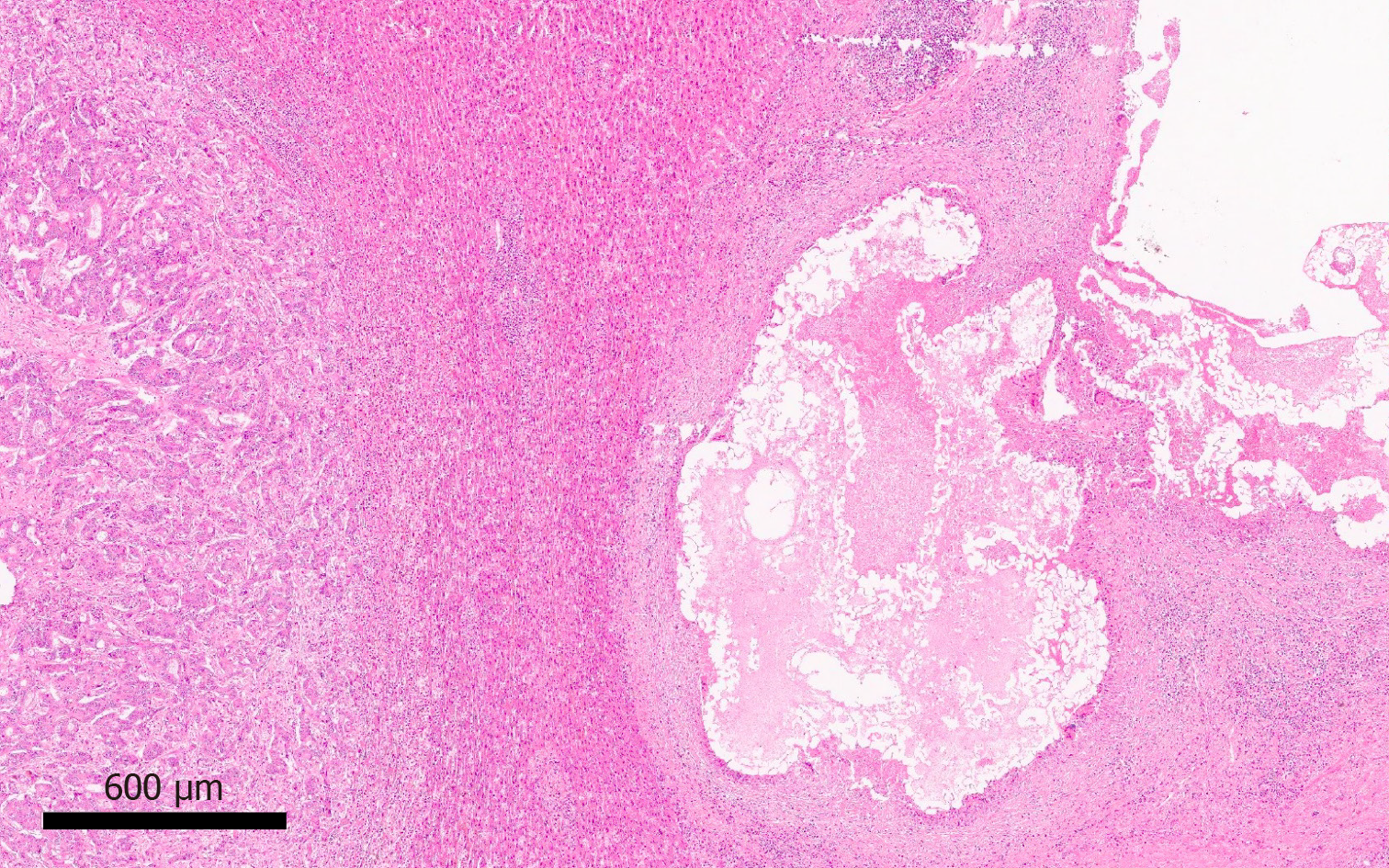
Figure 6 Embolic material within a large portal vein branch, with adjacent adenocarcinoma, in a patient who underwent preoperative portal vein embolization (Hematoxylin-eosin staining, × 4 magnification).
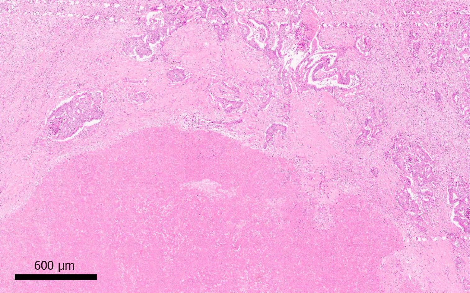
Figure 7 Extensive confluent tumour necrosis with fibrosis in keeping with a partial response to neoadjuvant therapy (Hematoxylin-eosin staining, × 4 magnification).
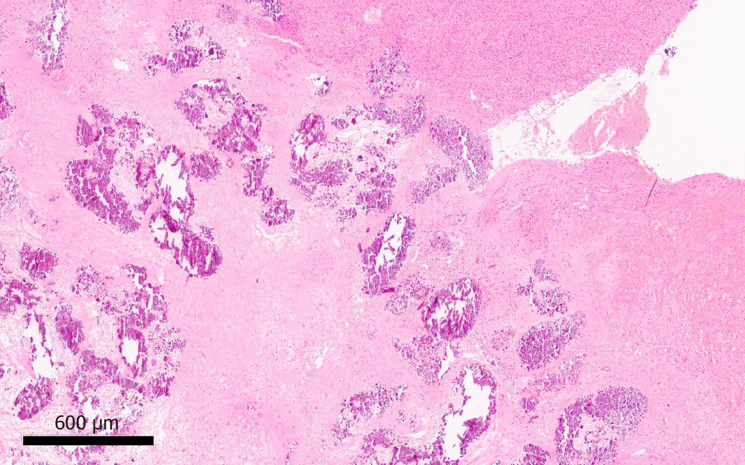
Figure 8 Confluent fibrosis and dystrophic calcification without viable residual tumour cells, consistent with pathological complete response to neoadjuvant therapy (Hematoxylin-eosin staining, × 4 magnification).
- Citation: Martin J, Petrillo A, Smyth EC, Shaida N, Khwaja S, Cheow H, Duckworth A, Heister P, Praseedom R, Jah A, Balakrishnan A, Harper S, Liau S, Kosmoliaptsis V, Huguet E. Colorectal liver metastases: Current management and future perspectives. World J Clin Oncol 2020; 11(10): 761-808
- URL: https://www.wjgnet.com/2218-4333/full/v11/i10/761.htm
- DOI: https://dx.doi.org/10.5306/wjco.v11.i10.761


