Copyright
©The Author(s) 2019.
World J Clin Oncol. Mar 24, 2019; 10(3): 161-165
Published online Mar 24, 2019. doi: 10.5306/wjco.v10.i3.161
Published online Mar 24, 2019. doi: 10.5306/wjco.v10.i3.161
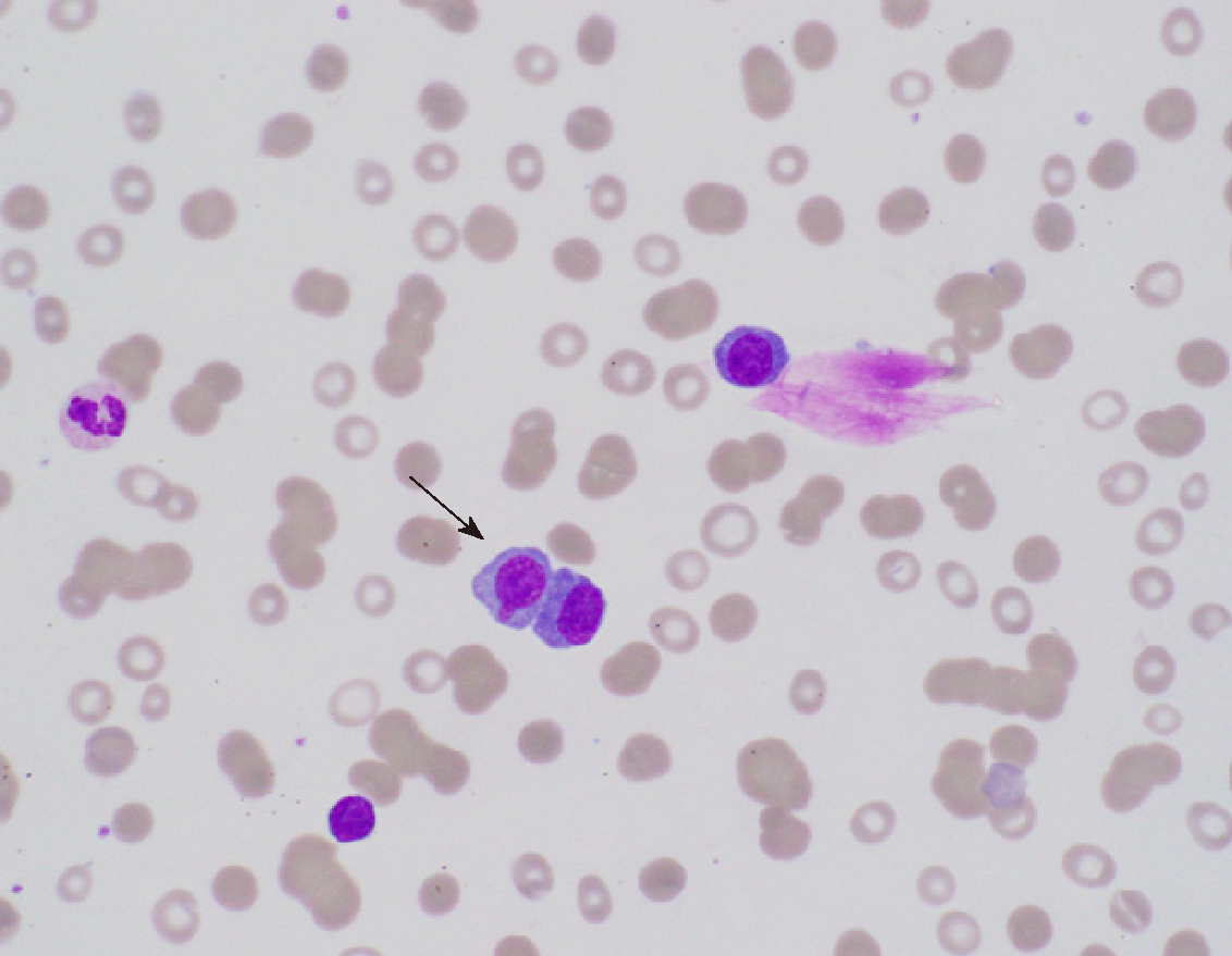
Figure 1 Peripheral blood smear.
Peripheral blood smear shows markedly increased plasma cells (black arrow), comprising approximately 20% of the white blood cells with eccentric, round to oval nuclei and basophilic cytoplasm.
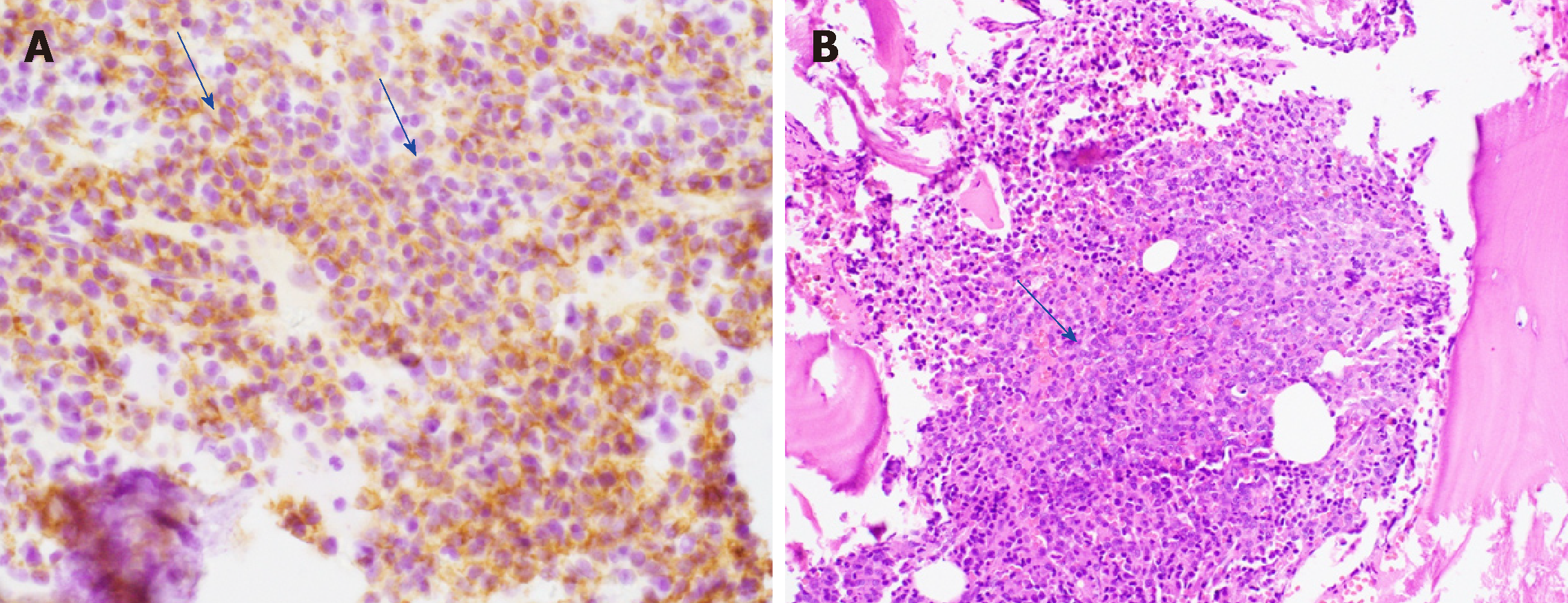
Figure 2 Microscopic examination of the bone marrow biopsy.
A: shows plasma cells (blue arrows) with CD138 immunohistochemical stain; B: extensive marrow replacement with neoplastic plasma cells (blue arrow).
- Citation: Jain AG, Faisal-Uddin M, Khan AK, Wazir M, Shen Q, Manoucheri M. Plasma cell leukemia - one in a million: A case report. World J Clin Oncol 2019; 10(3): 161-165
- URL: https://www.wjgnet.com/2218-4333/full/v10/i3/161.htm
- DOI: https://dx.doi.org/10.5306/wjco.v10.i3.161


