Copyright
©2010 Baishideng Publishing Group Co.
World J Clin Oncol. Nov 10, 2010; 1(1): 35-40
Published online Nov 10, 2010. doi: 10.5306/wjco.v1.i1.35
Published online Nov 10, 2010. doi: 10.5306/wjco.v1.i1.35
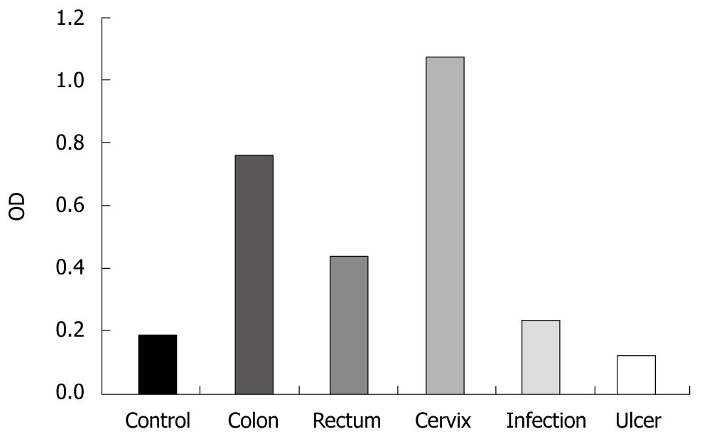
Figure 1 Level of anti-procathepsin D autoantibodies in patients with three types of cancer and two cancer-unrelated diseases evaluated by enzyme-linked immunosorbent assay.
Data represent mean value from 5 patients/group.
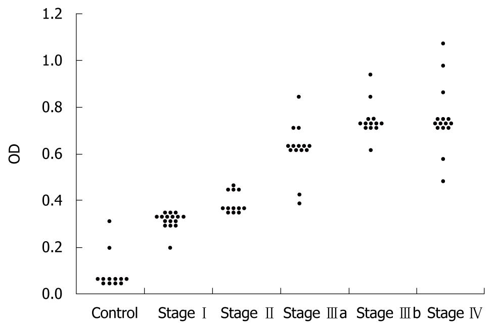
Figure 2 Level of anti-procathepsin D autoantibodies in patients with various stages of breast cancer evaluated by enzyme-linked immunosorbent assay.
Average age of patients in each group varied from 31 to 69.
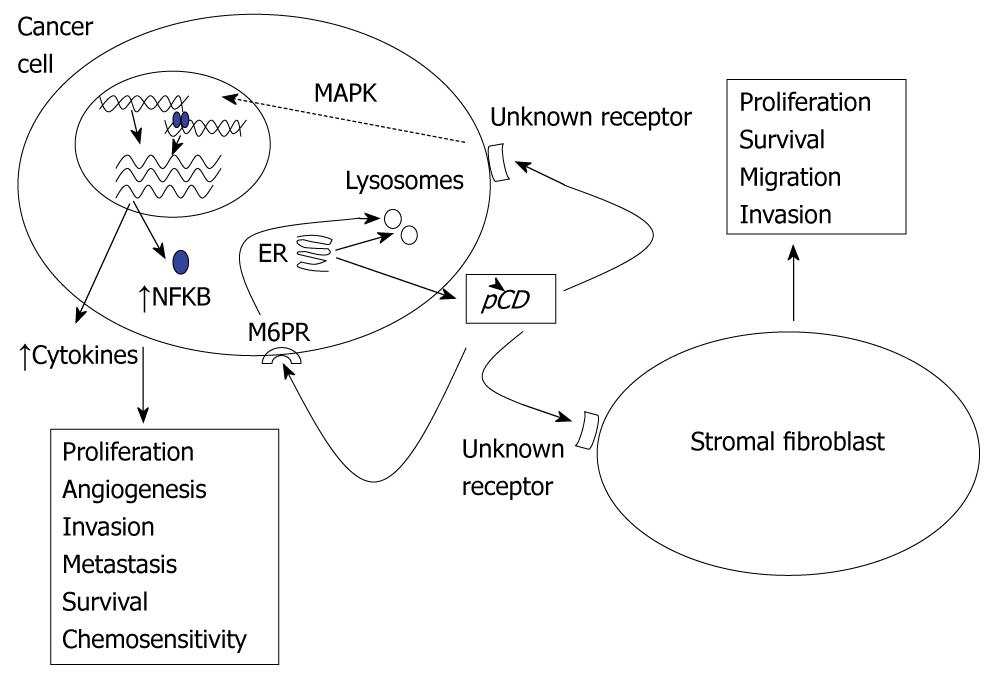
Figure 3 Proposed mechanism of procathepsin D/cathepsin D function in cancer progression.
The over-expressed procathepsin D (pCD) escapes the physiological intracellular targeting pathways and is secreted by the cancer cells. Small part of secreted pCD is bound and internalized via M6PR pathway, the rest by a yet unidentified receptor. The receptor-pCD interaction activates mitogen-activated protein kinases (MAPK) pathway with subsequent changes in expression of numerous cancer-promoting genes including NFKB2 and some cytokines. Interaction of pCD with endothelial and stromal cells is also involved.
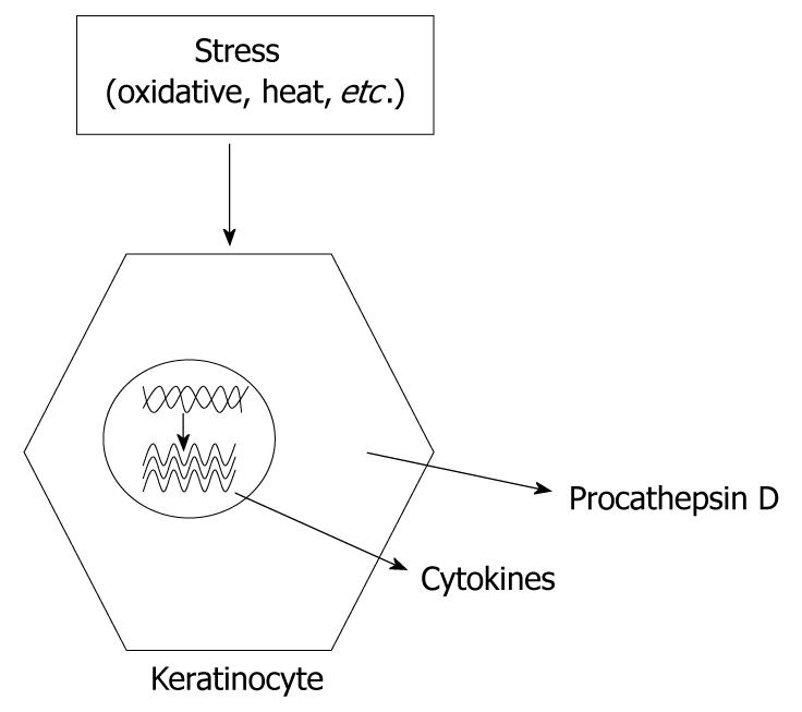
Figure 4 Stress affects keratinocytes resulting in increased both cytokine and procathepsin D synthesis and secretion leading to the elevated proliferation of keratinocytes.
- Citation: Vetvicka V, Vashishta A, Saraswat-Ohri S, Vetvickova J. Procathepsin D and cancer: From molecular biology to clinical applications. World J Clin Oncol 2010; 1(1): 35-40
- URL: https://www.wjgnet.com/2218-4333/full/v1/i1/35.htm
- DOI: https://dx.doi.org/10.5306/wjco.v1.i1.35


