Copyright
©The Author(s) 2017.
World J Gastrointest Pharmacol Ther. Feb 6, 2017; 8(1): 10-25
Published online Feb 6, 2017. doi: 10.4292/wjgpt.v8.i1.10
Published online Feb 6, 2017. doi: 10.4292/wjgpt.v8.i1.10
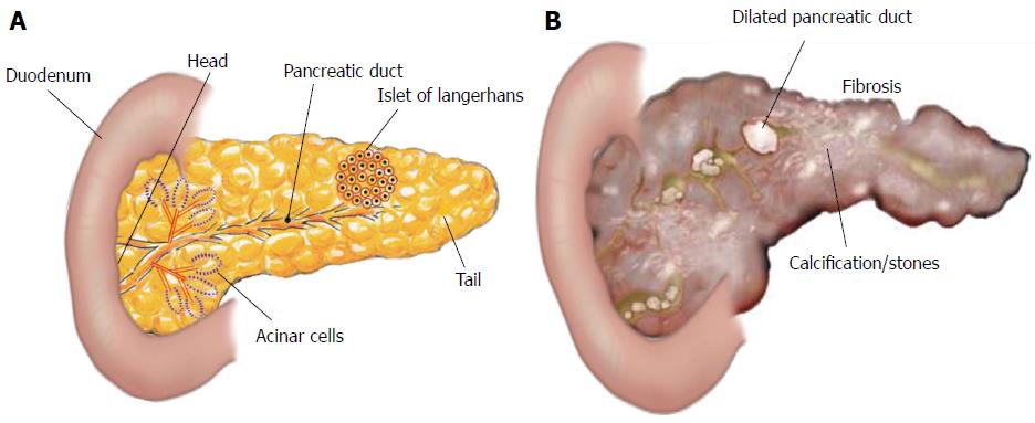
Figure 1 Structure of pancreas.
A: The pancreas is a leaf-like structure and has two types of cells: Exocrine cells, that include acinar pancreatic duct cells, and endocrine cells, that include islets of Langerhans; B: The inflammatory process in the pancreas promotes fibrosis (scarring of tissue), calcifications or stones, and dilated pancreatic duct.
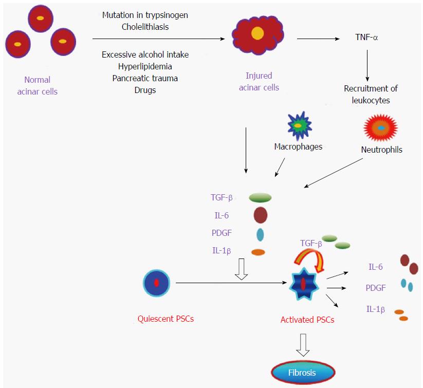
Figure 2 Pathogenesis of pancreatitis.
Diagrammatic representation of the onset of pancreatitis by damaged pancreatic acinar cells which in turn activates quiescent pancreatic stellate cells (PSCs) to become activated PSCs and promote subsequent fibrosis of pancreas. TNF-α: Tumor necrosis factor-α; TGF-β: Transforming growth factor-β; PDGF: Platelet-derived growth factor; IL: Interleukin.
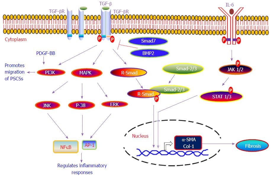
Figure 3 Various signaling pathways involved in the development of pancreatic fibrosis.
Diagrammatic representation of various molecular signaling pathways which are involved in the development of pancreatic fibrosis. TGF-β: Transforming growth factor-β; PDGF: Platelet-derived growth factor; IL: Interleukin; MAPK: Mitogen-activated protein kinase; PI3K: Phosphatidylinositol 3 kinase; AP-1: Activator protein-1; NFκB: Nuclear factor kappa B.
- Citation: Manohar M, Verma AK, Venkateshaiah SU, Sanders NL, Mishra A. Pathogenic mechanisms of pancreatitis. World J Gastrointest Pharmacol Ther 2017; 8(1): 10-25
- URL: https://www.wjgnet.com/2150-5349/full/v8/i1/10.htm
- DOI: https://dx.doi.org/10.4292/wjgpt.v8.i1.10


