Copyright
©The Author(s) 2016.
World J Gastrointest Pharmacol Ther. Nov 6, 2016; 7(4): 503-512
Published online Nov 6, 2016. doi: 10.4292/wjgpt.v7.i4.503
Published online Nov 6, 2016. doi: 10.4292/wjgpt.v7.i4.503
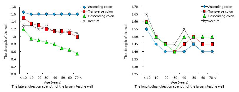
Figure 1 Change of strength with aging of the wall of the large intestine.
The descending colon (lateral direction) shows reduction in strength related to aging. In contrast, the ascending colon shows no reduction in strength related to aging.
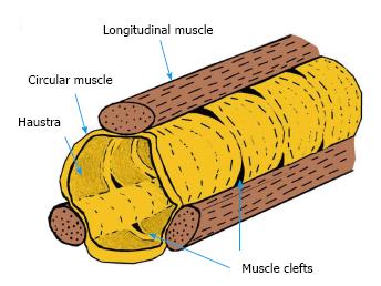
Figure 2 Muscle clefts.
Figure 3 According to Bernoulli's principle, the pressure at the extended site is increased.
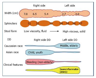
Figure 4 Distinctions in the background and symptoms of right and left diverticular disease.
The width of the right colon is about twice that of the left colon[53]. The large intestine contains seven sphincters[53], including S1: Busi ring; S2: Hirsh ring; S3: Cannon ring; S4: Payr-Straus ring; S5: Balli ring; S6: Moultier ring; S7: Rossi ring. Blue-colored areas represent the frequency at the site. In Caucasians, diverticular disease (DD) most frequently occurs on the left side and after middle age. In Asians, DD most frequently occurs on the right side and during childhood. Right-side diverticula is more likely to bleed but less likely to develop the severe diverticula-associated complications of perforation, abscess formation, fistulation, or structuring[7,54].
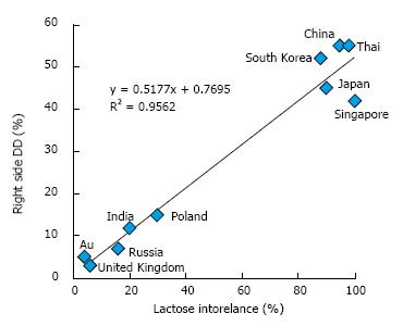
Figure 5 Relationship between lactose intolerance and the right-side diverticular disease.
Au: Australia; DD: Diverticular disease.
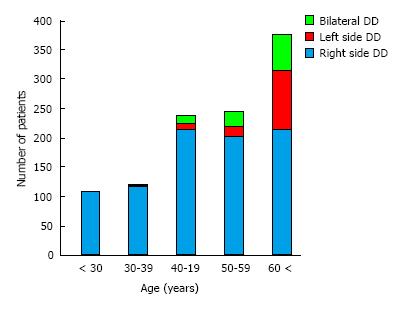
Figure 6 Relationship between the site of and age at onset for diverticular disease.
DD: Diverticular disease.
- Citation: Uno Y, van Velkinburgh JC. Logical hypothesis: Low FODMAP diet to prevent diverticulitis. World J Gastrointest Pharmacol Ther 2016; 7(4): 503-512
- URL: https://www.wjgnet.com/2150-5349/full/v7/i4/503.htm
- DOI: https://dx.doi.org/10.4292/wjgpt.v7.i4.503


