Copyright
©The Author(s) 2015.
World J Gastrointest Pharmacol Ther. Nov 6, 2015; 6(4): 223-237
Published online Nov 6, 2015. doi: 10.4292/wjgpt.v6.i4.223
Published online Nov 6, 2015. doi: 10.4292/wjgpt.v6.i4.223
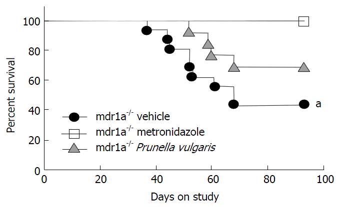
Figure 1 Effect of treatment with a Prunella vulgaris ethanolic extract on the onset of colitis in mdr1a-/- mice.
Mdr1a-/- mice were removed from the study as they developed severe clinical disease (e.g., > 15% weight loss) before the termination of the experiment as described in Materials and Methods. aP < 0.05, as compared to FVBWT control mice. Vehicle-treated mdr1a-/- mice n = 16, metronidazole-treated mdr1a-/- mice n = 10, Prunella vulgaris-treated mdr1a-/- mice n = 13. This survival (i.e., mice remaining on study) curve is representative of two independent experiments.
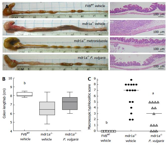
Figure 2 Oral administration of a Prunella vulgaris extract attenuated both microscopic and macroscopic cecal lesions in mdr1a-/- mice.
A: Representative photographs of ceca and colons (left) and representative photomicrographs (200 ×) of histological sections of ceca (right) collected at necropsy from FVBWT or mdr1a-/- mice treated with either vehicle or Prunella vulgaris (P. vulgaris) extract; B: Colon lengths were measured at necropsy and the group range is represented. Whiskers indicate minimum and maximum values, while the horizontal line represents the group median; C: Macroscopic typhlocolitic scores were assigned at necropsy as described in the Materials and Methods (Max/Severe = 9, Min/Healthy = 0). aP < 0.05, bP < 0.01 compared to mdr1a-/- vehicle as calculated by Kruskal-Wallis test. Vehicle-treated FVBWT mice n = 6, vehicle-treated mdr1a-/- mice n = 16, P. vulgaris-treated mdr1a-/- mice n = 13.
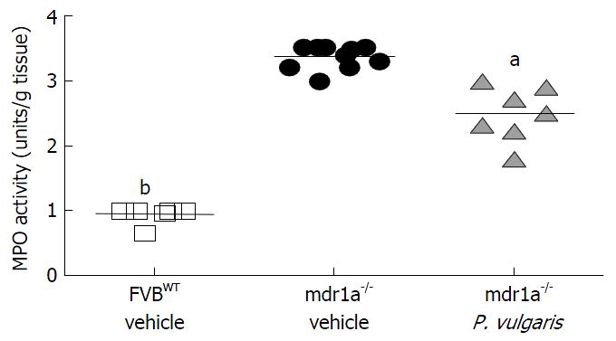
Figure 3 Administration of a Prunella vulgaris ethanolic extract reduced local myeloperoxidase activity in the colon of mdr1a-/- mice.
Homogenates of colonic tissue were subjected to an assay for MPO activity. aP < 0.05, bP < 0.01 compared to mdr1a-/- vehicle as calculated by Kruskal-Wallis test. Vehicle-treated FVBWT mice n = 6, vehicle-treated mdr1a-/- mice n = 10, P. vulgaris-treated mdr1a-/- mice n = 7. MPO: Myeloperoxidase; P. vulgaris: Prunella vulgaris.
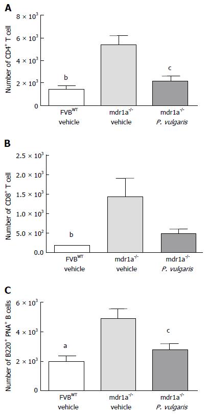
Figure 4 Evaluation of T cell and B cell subsets in the cecal tonsil of mdr1a-/- mice treated with prunella vulgaris ethanolic extract.
Cecal tonsils were excised at necropsy, single cell suspensions prepared and labeled for flow cytometric analysis as described in Materials and Methods. Absolute numbers of (A) CD4+ T cells; (B) CD8ß+ T cells; and (C) B220+PNA+ germinal center B cells in the cecal tonsils of mice. aP < 0.05, bP < 0.01 compared to mdr1a-/- vehicle as calculated by Kruskal-Wallis test. cP < 0.05 compared to mdr1a-/- vehicle as calculated by unpaired t-test. The n for each group is equal to that noted in Figure 1 and data are representative of two independent experiments. Vehicle-treated FVBWT mice n = 5-6, vehicle-treated mdr1a-/- mice n = 8-10, P. vulgaris-treated mdr1a-/- mice n = 5-7.
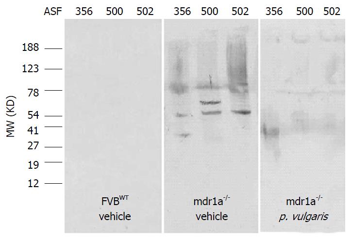
Figure 5 Vehicle-treated mdr1a-/- mice with severe colitic inflammation developed serum antibody to antigens derived from gut microbiota while those treated with prunella vulgaris extract do not.
Whole cell sonicates of three separate clostridial species present as part of the intestinal microbiota (altered Schaedler flora members 356, 500 and 502) were subjected to SDS-PAGE. Western blot analysis was performed using sera collected at necropsy. Antigens in lanes represented in each panel are as follows from left to right: ASF502, ASF500 and ASF356.
-
Citation: Haarberg KM, Wymore Brand MJ, Overstreet AMC, Hauck CC, Murphy PA, Hostetter JM, Ramer-Tait AE, Wannemuehler MJ. Orally administered extract from
Prunella vulgaris attenuates spontaneous colitis in mdr1a-/- mice. World J Gastrointest Pharmacol Ther 2015; 6(4): 223-237 - URL: https://www.wjgnet.com/2150-5349/full/v6/i4/223.htm
- DOI: https://dx.doi.org/10.4292/wjgpt.v6.i4.223


