Copyright
©The Author(s) 2015.
World J Gastrointest Pharmacol Ther. Nov 6, 2015; 6(4): 145-155
Published online Nov 6, 2015. doi: 10.4292/wjgpt.v6.i4.145
Published online Nov 6, 2015. doi: 10.4292/wjgpt.v6.i4.145
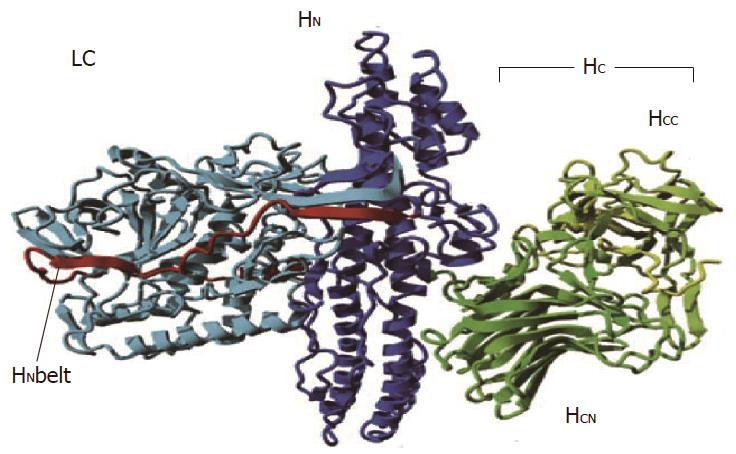
Figure 1 Structure of botulinum toxin A.
The Cα backbone is represented as ribbons with the LC in cyan, the HN in dark blue and the HC in a green to yellow gradient highlighting the HCN and HCC subdomains. The HN belt is in red. HN + HC: 100 kDa heavy chain; HN: 50 kDa amino terminus; HC: 50 kDa carboxy terminal of the heavy chain; HCC: β-beta tree foil fold heavy chain subdomain; HCN: β-sheet jelly roll fold heavy chain subdomain; LC: Light chain. Adapted with permission from Montal[17].
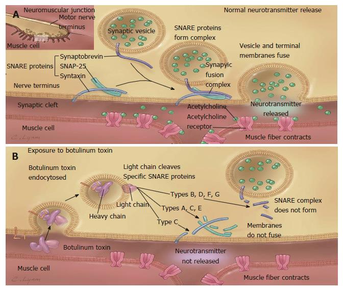
Figure 2 Mechanism of action of botulinum neurotoxin.
A: Release of acetylcholine at the neuromuscular junction is mediated by the assembly of a synaptic fusion complex that allows the membrane of the synaptic vesicle containing acetylcholine to fuse with the neuronal cell membrane. The synaptic fusion complex is a set of SNARE proteins, which include synaptobrevin, SNAP-25, and syntaxin. After membrane fusion, acetylcholine is released into the synaptic cleft and then bound by receptors on the muscle cell; B: Botulinum toxin binds to the neuronal cell membrane at the nerve terminus and enters the neuron by endocytosis. The light chain of botulinum toxin cleaves specific sites on the SNARE proteins, preventing complete assembly of the synaptic fusion complex and thereby blocking acetylcholine release. Botulinum toxins types B, D, F, and G cleave synaptobrevin; types A, C, and E cleave SNAP-25; and type C cleaves syntaxin. Without acetylcholine release, the muscle is unable to contract. SNARE: Soluble NSF-attachment protein receptor; NSF: N-ethylmaleimide-sensitive fusion protein; SNAP-25: Synaptosomal-associated protein of 25 kD. Reproduced with permission from Arnon et al[91].
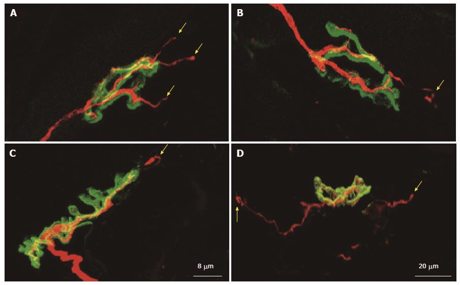
Figure 3 Neuronal sprouting and remodeling of the neuromuscular junction.
Remodeling of the neuromuscular junction in extensor digitorum longus muscle at 10 (A-C) and 21 d (D) after a single injection of botulinum neurotoxin type C. Axons and nerve terminals were immunolabelled (red). To localize the junctions, nicotinic acetylcholine receptors were stained (green). Note the sprouts that emerge from the original motor endplate and project along muscle fibers (yellow arrows). Reproduced with permission from Morbiato et al[92].
- Citation: Nassri A, Ramzan Z. Pharmacotherapy for the management of achalasia: Current status, challenges and future directions. World J Gastrointest Pharmacol Ther 2015; 6(4): 145-155
- URL: https://www.wjgnet.com/2150-5349/full/v6/i4/145.htm
- DOI: https://dx.doi.org/10.4292/wjgpt.v6.i4.145


