Copyright
©2014 Baishideng Publishing Group Inc.
World J Gastrointest Pharmacol Ther. Nov 6, 2014; 5(4): 200-208
Published online Nov 6, 2014. doi: 10.4292/wjgpt.v5.i4.200
Published online Nov 6, 2014. doi: 10.4292/wjgpt.v5.i4.200
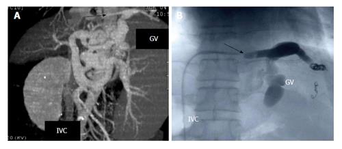
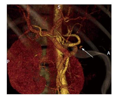
Figure 1 Balloon retrograde transvenous obliteration[59].
A: Multidimensional computed tomography revealing varices supplied by the left gastric vein (GV) and subsequent drainage into the subphrenic vein (illustrated by the arrow) which is connected to the inferior vena cava (IVC); B: Fluoroscopic image of B-RTO depicting placement of the occlusive balloon catheter into the bleeding vessel (demonstrated by the arrow). B-RTO: Balloon-occluded retrograde transvenous obliteration.
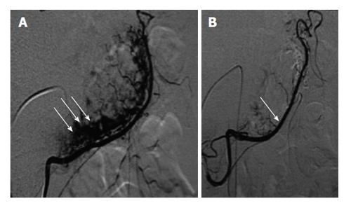
Figure 2 Selective transarterial embolization[60].
A: Selective catheterization of the right gastro epiploic artery in the setting of angiodysplasia. The arrows depict the pathologic vessels at the greater curvature of the stomach; B: Post embolization angiogram revealing control of the bleeding vessel.
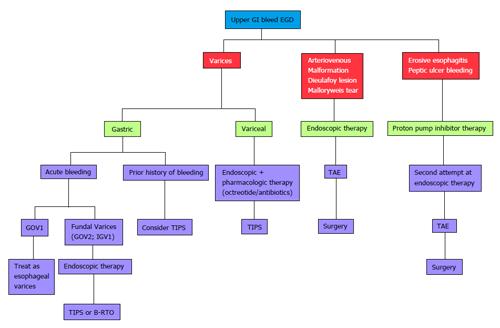
Figure 3 Algorithmic approach to upper gastrointestinal bleeding.
EGD: Esophagoduodeonscopy; TAE: Transcatheter arterial embolization; TIPS: Transjugular intrahepatic portosystemic shunt; GOV1: Gastroesophageal Varices 1; B-RTO: Balloon-occluded retrograde transvenous obliteration; GI: Gastrointestinal; IGV: Isolated gastric varices.
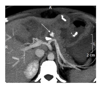
Figure 4 Unenhanced computed tomography showing contrast media extravasation anterior to the splenic artery[61].
Figure 5 Multiphasic multidetector computed topography angiography (arterial phase) showing extravasation anterior to the splenic artery[61].
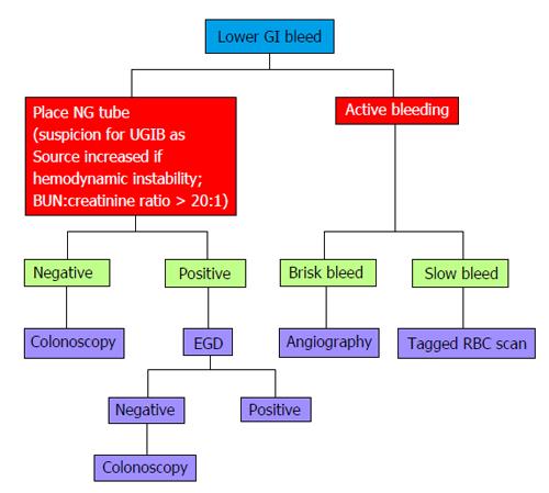
Figure 6 Algorithmic approach to lower gastrointestinal bleeding.
EGD: Esophagoduodeonscopy; GI: Gastrointestinal; NG: Nasogastric; BUN: Blood urea nitrogen; RBC: Red blood cell; UGIB: Upper gastrointestinal bleeding.
- Citation: Parekh PJ, Buerlein RC, Shams R, Vingan H, Johnson DA. Evaluation of gastrointestinal bleeding: Update of current radiologic strategies. World J Gastrointest Pharmacol Ther 2014; 5(4): 200-208
- URL: https://www.wjgnet.com/2150-5349/full/v5/i4/200.htm
- DOI: https://dx.doi.org/10.4292/wjgpt.v5.i4.200


