Copyright
©The Author(s) 2017.
World J Gastrointest Pathophysiol. May 15, 2017; 8(2): 39-50
Published online May 15, 2017. doi: 10.4291/wjgp.v8.i2.39
Published online May 15, 2017. doi: 10.4291/wjgp.v8.i2.39
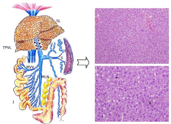
Figure 1 Liver histopathological changes of a rat after three months with triple partial portal vein ligation.
Hepatic steatotic areas are mainly distributed in zone 1 of the liver acinus, but tipically hepatocyte ballooning is more apparent near zone 3. Both macrovesicular and microvesicular steatosis are evident as well as scattered necroinflammatory foci (arrowheads) (H and E stain, × 200). I: Intestine; MVV: Mesenteric venous vasculopathy; S: Spleen; SL: Steatotic liver; TPVL: Triple partial portal vein ligation.
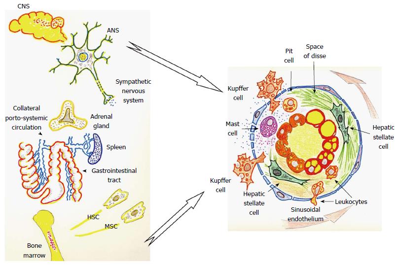
Figure 2 Inflammatory phenotypes, neurogenic and immune in prehepatic portal hypertensive rat.
ANS: Autonomic nervous system; CNS: Central nervous system; HSC: Hematological stem cell; MSC: Mesenchymal stem cell.
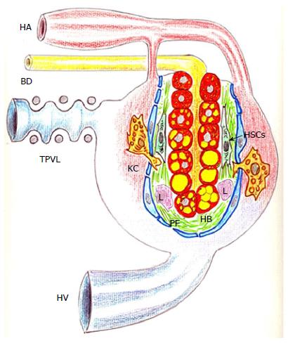
Figure 3 Schematic representation of the liver parenchyma after partial portal vein in the rat.
The deposit of lipids within the hepatocytes and the liver arterialization, associated with defenestration of the sinusoidal endothelium, and the perisinuosidal fibrosis stand out. BD: Bile duct; HA: Hepatic artery; HV: Hepatic vein; HB: Hepatocyte ballooning; HSCs: Hepatic stellate cells; KC: Kupffer cell; L: Leukocyte; PF: Perisinuosidal fibrosis; TPVL: Triple partial portal vein ligation.
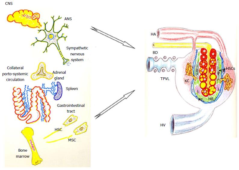
Figure 4 The preferential expression of the neurogenic and immune inflammatory phenotypes into the space of Disse induces hepatic steatosis, steatohepatitis and fibrosis in portal hypertensive rats by triple partial portal vein ligation.
ANS: Autonomic nervous system; BD: Bile duct; CNS: Central nervous system; HA: Hepatic artery; HB: Hepatocyte ballooning; HSC: Hematological stem cell; HSCs: Hepatic stellate cell; HV: Hepatic vein; KC: Kupffer cell; L: Leukocyte; MSC: Mesenchymal stem cell; PF: Perisinuosidal fibrosis; TPVL: Triple partial portal vein ligation.
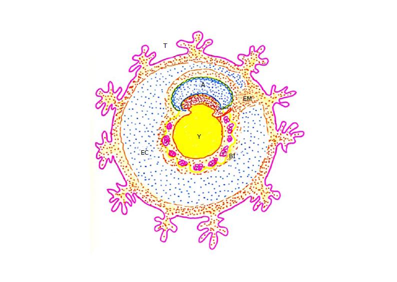
Figure 5 Schematic representation of early embryo development.
The extra-embryonic mesoderm and the exocoelomic cavity connect the trophoblast with the amnion and the yolk sac. A: Amnion; BI: Blood islands; EC: Exocoelomic cavity; EM: Extra-embryonic mesoderm; T: Trophoblast; Y: Yolk sac.
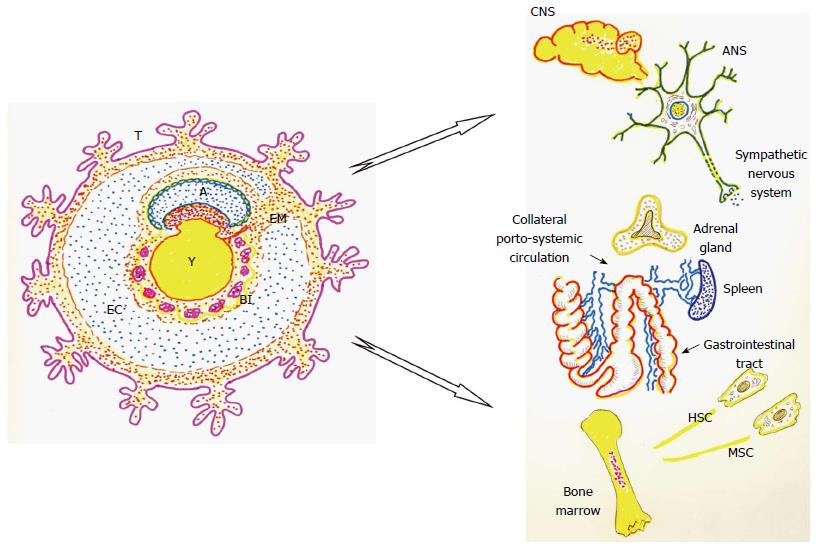
Figure 6 The coelomic-amniotic functions could be hypothetically recapitulated by the systemic neurogenic phenotype activation, while the trophoblastic-yolk sac functions could be carried out by the systemic immune inflammatory phenotype activation.
A: Amnion; ANS: Autonomic nervous system; BI: Blood islands; CNS: Central nervous system; EC: Exocoelomic cavity; EM: Extra-embryonic mesoderm; HSC: Hematological stem cell; MSC: Mesenchymal stem cell; T: Trophoblast; Y: Yolk sac.
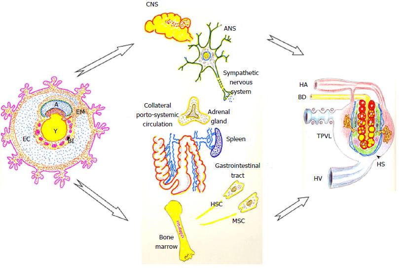
Figure 7 The recapitulated systemic extra-embryonic functions, that is, the coelomic-amniotic and the trophoblastic-yolk sac (or vitellum), are located into the space of Disse.
This fact results in the development of hepatic steatosis and steatohepatitis in prehepatic portal hypertensive rats. A: Amnion; ANS: Autonomic nervous system; BD: Bile duct; BI: Blood islands; CNS: Central nervous system; EM: Extraembryonic mesoderm; HA: Hepatic artery; HS: Steatotic liver; HSC: Hematological stem cell; HV: Hepatic vein; MSC: Mesenchymal stem cell; TPVL: Triple partial portal vein ligation; Y: Yolk sac.
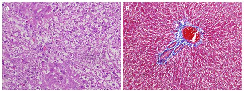
Figure 8 Histological appearance of non-alcoholic steatohepatitis in portal hypertensive rats at three months of postoperative evolution.
It’s shown the hepatocyte ballooning (A) with lobular inflammation and perisinuosidal fibrosis (B) (H and E stain, × 200).
- Citation: Aller MA, Arias N, Peral I, García-Higarza S, Arias JL, Arias J. Embrionary way to create a fatty liver in portal hypertension. World J Gastrointest Pathophysiol 2017; 8(2): 39-50
- URL: https://www.wjgnet.com/2150-5330/full/v8/i2/39.htm
- DOI: https://dx.doi.org/10.4291/wjgp.v8.i2.39


