Copyright
©2014 Baishideng Publishing Group Inc.
World J Gastrointest Pathophysiol. Aug 15, 2014; 5(3): 188-199
Published online Aug 15, 2014. doi: 10.4291/wjgp.v5.i3.188
Published online Aug 15, 2014. doi: 10.4291/wjgp.v5.i3.188
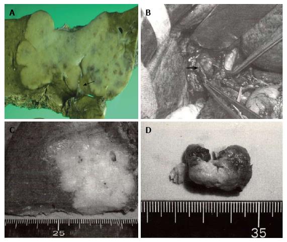
Figure 1 Some intraductal growth-type intrahepatic cholangiocarcinomas are considered to be an intraductal papillary neoplasm of the bile duct, this classification system provides useful information during surgery.
A: Gross feature of mass-forming (MF) + periductal infiltrating (PI)-type intrahepatic cholangiocarcinoma (ICC) obtained by left hepatectomy with bile duct resection. The carcinoma spreads along the hilar biliary tree (arrow) in communication with a white firm mass; B-D: Operative findings and resected specimens of MF + intraductal growth (IG)-type ICC. The common hepatic duct is incised, and the soft tumor comprising the intraductal components is easily removed (arrow) without infiltration to the ductal wall; C: Hepatic anterior segmentectomy is performed for MF components; D: IG components are composed of tan-colored soft tissues with necrosis.
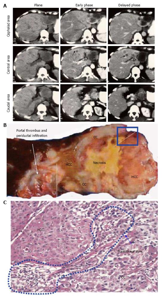
Figure 2 Case presentation of combined hepatocellular-cholangiocarcinoma.
A: Preoperative computed tomography shows a large mass composed of two major components that replaces the lateral segment. The mass shows ringed enhancement in the delayed phase in the cephalad area and early enhancement with washout in the delayed phase in the caudal area. Intrahepatic metastases are observed in the S4 segment (arrow); B: Gross features of the resected specimen. Hepatocellular carcinoma (HCC) components are composed of tan-colored soft tissues. Cholangiocarcinoma (CC) components are composed of white firm tissues with central necrosis; C: Small round cells and fibrous stroma are observed at the boundary area between the HCC and CC components (blue flame in panel B).
Figure 3 Resected case of cholangiocellular carcinoma.
A: Computed tomography (CT) reveals a mass showing ringed enhancement with portal venous penetration; B: CT findings reflect the non-infiltrative growth of the tumor to the portal tract; C: Histopathologically, the size of the carcinoma cells is small, with the cells forming anastomosing patterns with abundant fibrous stroma.
- Citation: Sanada Y, Kawashita Y, Okada S, Azuma T, Matsuo S. Review to better understand the macroscopic subtypes and histogenesis of intrahepatic cholangiocarcinoma. World J Gastrointest Pathophysiol 2014; 5(3): 188-199
- URL: https://www.wjgnet.com/2150-5330/full/v5/i3/188.htm
- DOI: https://dx.doi.org/10.4291/wjgp.v5.i3.188


