Copyright
©The Author(s) 2020.
World J Gastrointest Pathophysiol. Mar 13, 2020; 11(1): 1-19
Published online Mar 13, 2020. doi: 10.4291/wjgp.v11.i1.1
Published online Mar 13, 2020. doi: 10.4291/wjgp.v11.i1.1
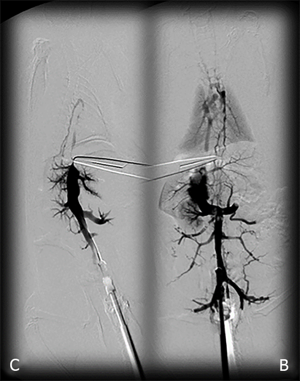
Figure 1 BPC 157 and venography assessment in rats with a suprahepatic occlusion of the inferior vena cava.
Inferior vena cava venography at the level of the bifurcation at 15 minutes ligation time. Blood flow through the inferior vena cava through the azygos vein to the superior vena cava, portal vein to superior-inferior mesenteric vein-rectal vein-left iliac vein-inferior vena cava in BPC 157-treated rats (B), but not in the controls (C).
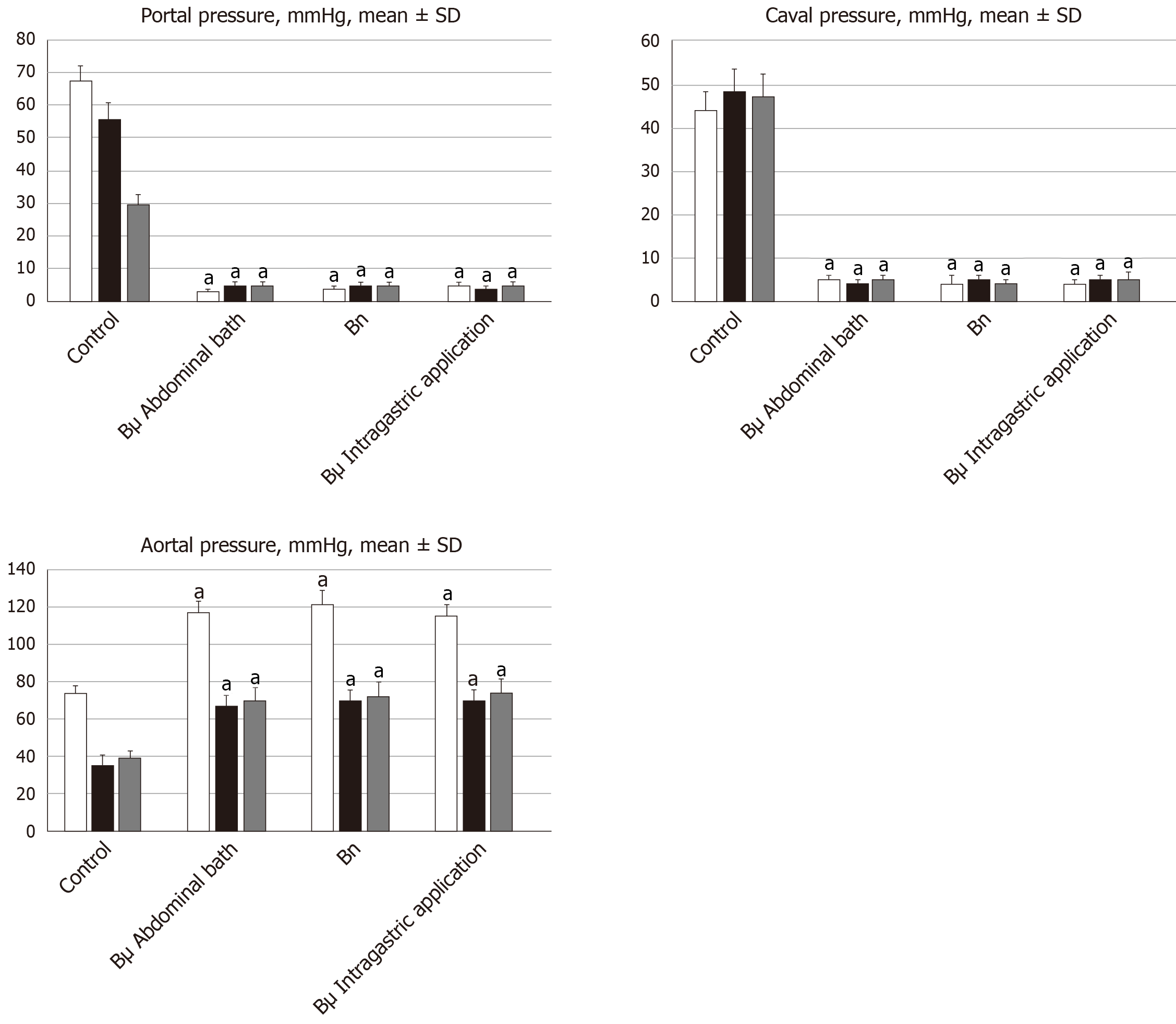
Figure 2 Antagonizing effect of BPC 157 on portal and caval hypertension, and aortal hypotension.
Portal, caval and aortal pressure (mmHg), mean ± SD. For portal vein, vena cava, and abdominal aorta pressure recordings, medication of BPC 157 [10 µg/kg (Bµg), 10 ng/kg (Bng)] or saline (5 mL/kg; control) was applied in rats intragastrically or as an abdominal bath, at 15 min (white bars), 24 h (black bars) or 48 h (gray bars) post-ligation. aP < 0.05 vs control.
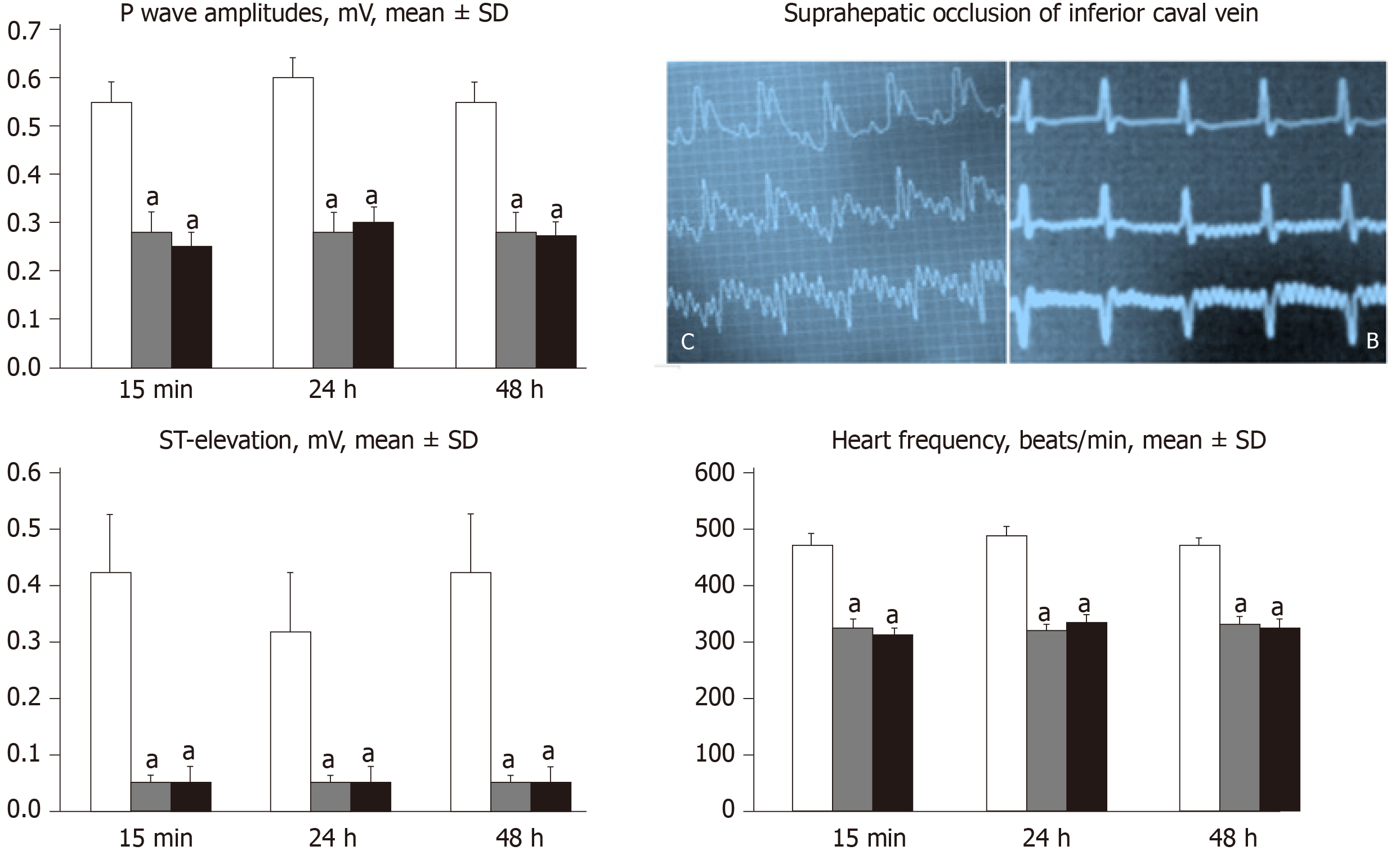
Figure 3 Counteracting effect of BPC 157 in rats with a suprahepatic occlusion of the inferior vena cava on peaked P waves, sinus tachycardia, ST-elevation 15 min post-ligation [controls (C), BPC 157 rats (B)] and assessment at 15 min, 24 h and 48 h ligation-time, mean ± SD.
Treatment with 10 µg/kg BPC 157 (gray bars), 10 ng/kg BPC 157 (black bars), or saline (1 mL/rat; white bars) as a bath administration given at 1 min post-ligation. aP < 0.05 vs control.
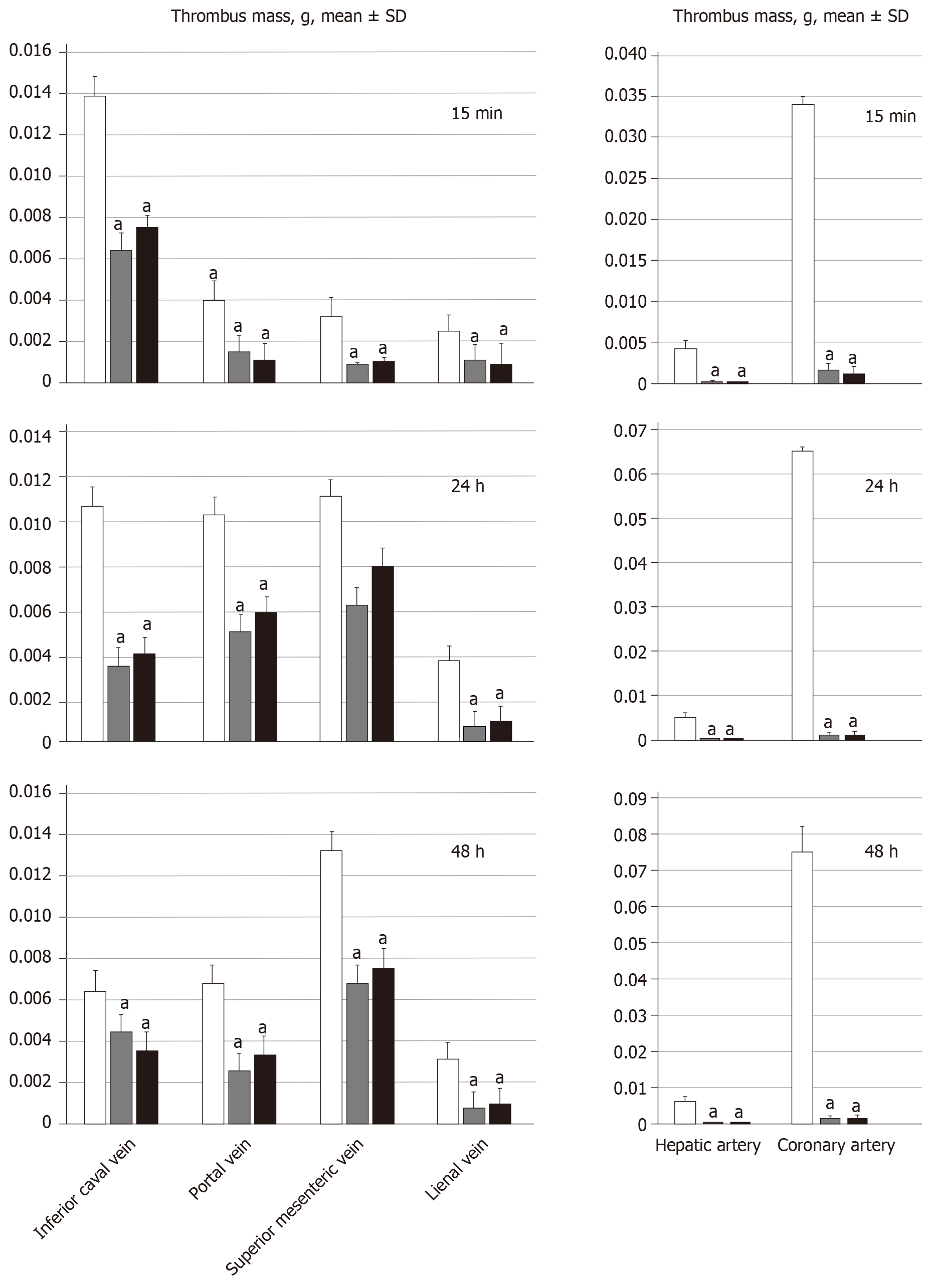
Figure 4 BPC 157 attenuates thrombus presentation (thrombus mass, g) in veins (inferior vena cava, portal vein, superior mesenteric vein, and splenic vein, SV) and arteries (hepatic artery and coronary artery) following suprahepatic occlusion of the inferior vena cava.
Assessment at 15 min, 24 h and 48 h post-ligation; mean ± SD. Treatment with 10 µg/kg BPC 157 (gray bars), 10 ng/kg BPC 157 (black bars), or saline (1 mL/rat; white bars) as a bath administration given at 1 min post-ligation. aP < 0.05 vs control.
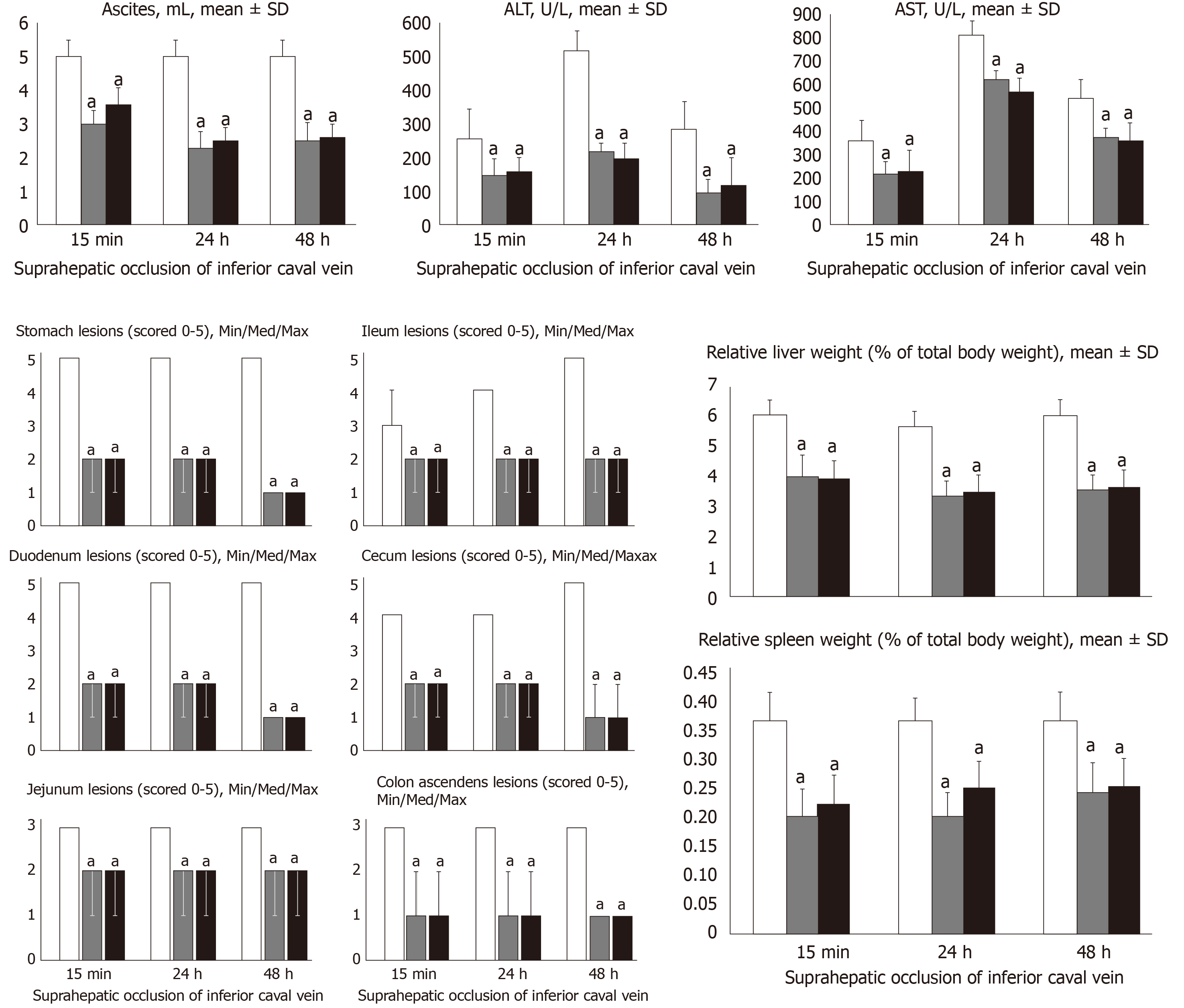
Figure 5 BPC 157 counteracts ascites (mL), serum enzyme levels (U/L), hepatomegaly and splenomegaly (mean ± SD), stomach, duodenal, jejunal, ileal, cecal and ascending colon lesions (scored 0–5; Min/Med/Max), following suprahepatic occlusion of the inferior vena cava.
Assessment at 15 min, 24 h and 48 h post-ligation. Treatment with 10 µg/kg BPC 157 (gray bars), 10 ng/kg BPC 157 (black bars), or saline (1 mL/rat; white bars) as a bath administration given at 1 min ligation. aP < 0.05 vs control.
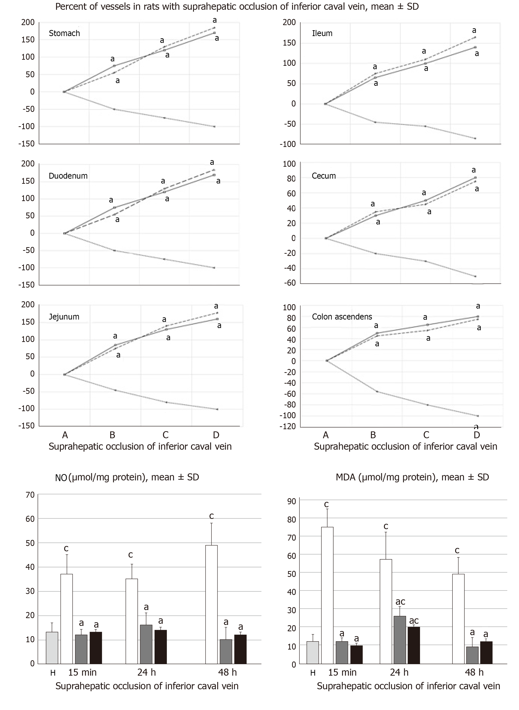
Figure 6 BPC 157 improves vessel presentation following suprahepatic occlusion of the inferior vena cava (upper), and eliminates or attenuates increased nitric oxide and malondialdehyde levels (nmol/mg protein) in liver.
Vessels were assessed as to whether they were (filled or cleared out at the stomach, and between the arcade vessels on the ventral side for 1 cm long segments of duodenum, jejunum and ascending colon, and between 10 vessels from the proximal to distal cecum. Assessment was made throughout the experiment, with the point (A) immediately before therapy considered to be 100%, at selected time points before (A) and after therapy (B, C and D) in rats with suprahepatic occlusion of the inferior vena cava at 5 (B), 10 (C) and 15 (D) minutes post-ligation (mean ± SD). The gross appearance of the tissue was recorded using a USB microscope camera (upper). Counteraction of the increased nitric oxide and malondialdehyde levels in liver (lower) at 15 min, 24 h and 48 h post-ligation. Treatment with 10 µg/kg BPC 157 (gary bars), 10 ng/kg BPC 157 (black bars), or saline (1 mL/rat; white bars) as a bath administration given at 1 min post-ligation. aP < 0.05 vs saline; cP < 0.05 vs healthy liver (H). NO: Nitric oxide; MDA: Malondialdehyde.
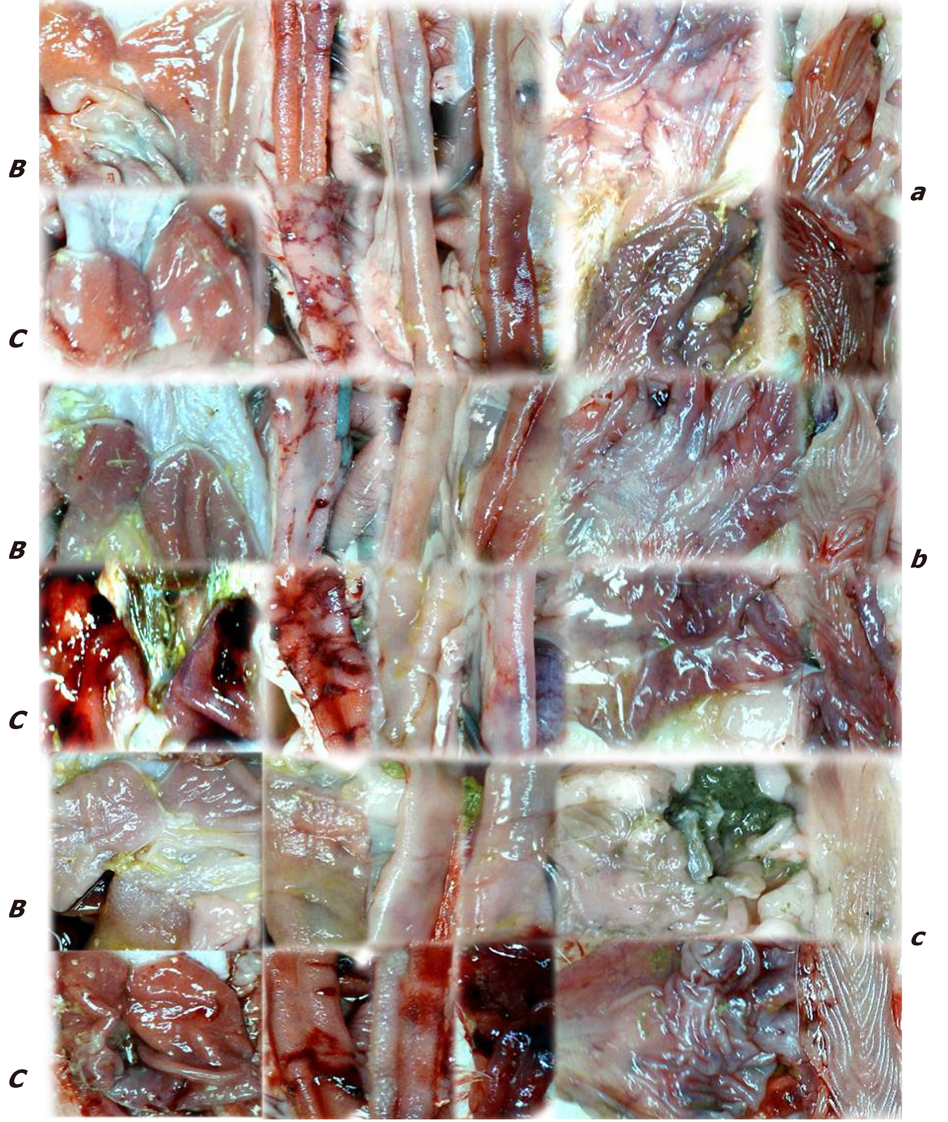
Figure 7 Gross presentation of lesions (C) and lesions attenuation (B) in rats with suprahepatic occlusion of inferior vena cava.
Lesions were assessed using a camera attached to a USB microscope (Veho Discovery Deluxe VMS-004), at 15 min (upper, a), 24 h (middle, b), and 48 h (low, c) post-ligation in the gastrointestinal tract. From left to right, the stomach, duodenum, jejunum, ileum, cecum and ascending colon on treatment with saline (C) or BPC 157 (B).
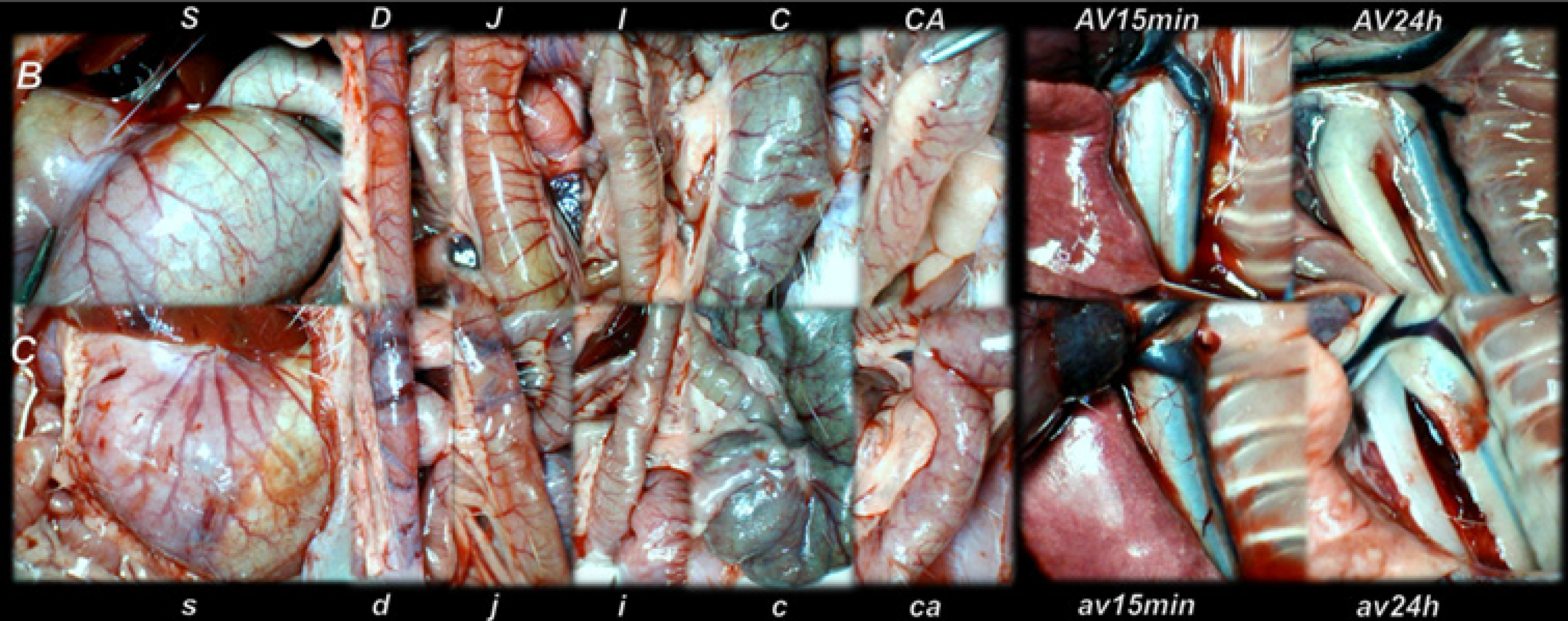
Figure 8 Gross vessel presentation in rats treated with saline (C, small letters, lower) and improvement in rats treated with BPC 157 (B, capitals, upper).
Assessments were made using a camera attached to a USB microscope (Veho Discovery Deluxe VMS-004) in the stomach (s/S), duodenum (d/D), jejunum (j/J), ileum (i/I), cecum (c/C) and ascending colon (ca/CA) of rats with suprahepatic occlusion of the inferior vena cava at 15 min post-ligation (left), and azygos vein presentation at 15 min (av15min/AV15min) and 24 h (av24h/AV24h) post-ligation (right).
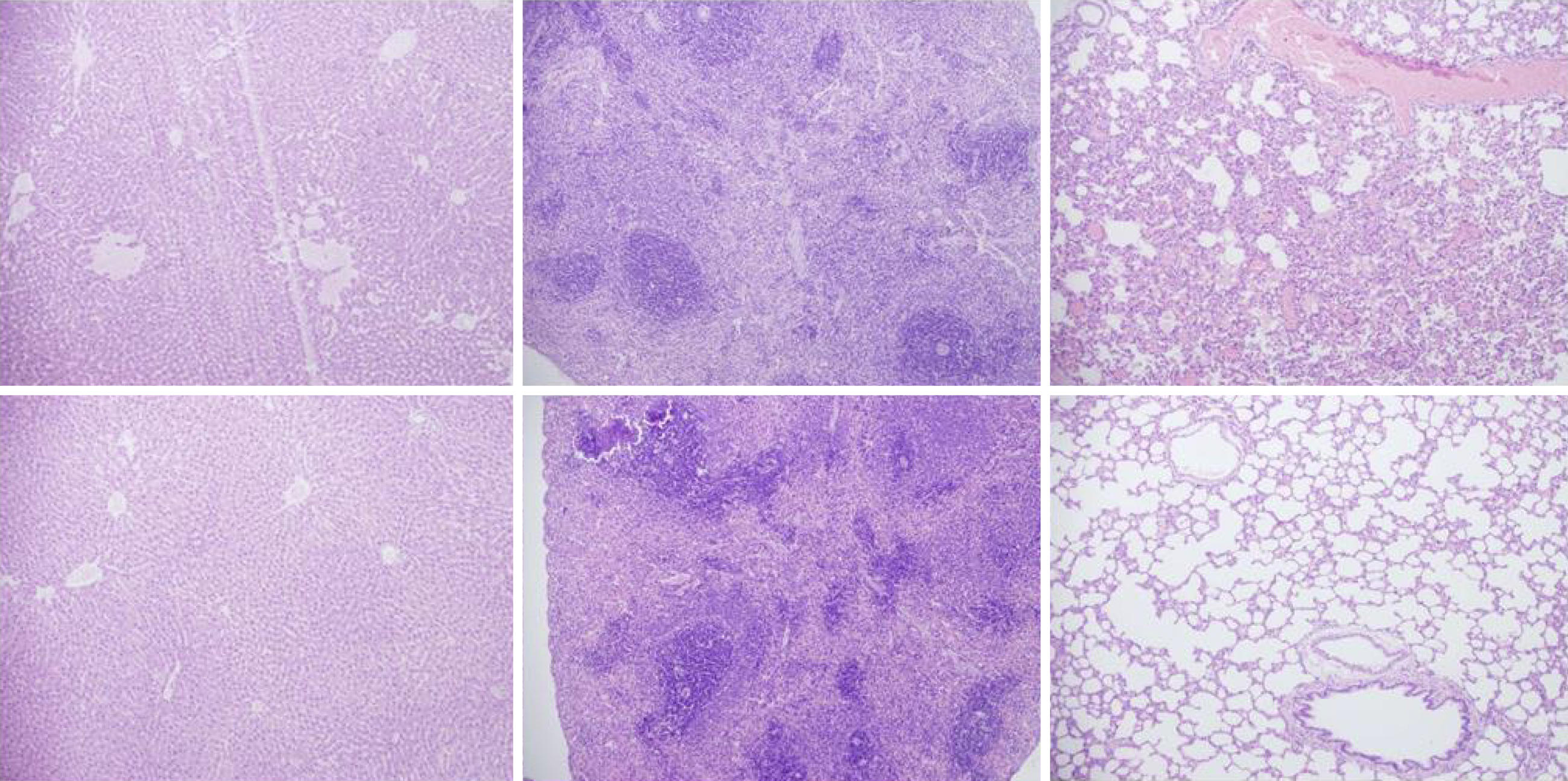
Figure 9 Histological presentation in rats with suprahepatic ligation of the inferior vena cava.
Assessment (stained with hematoxylin-eosin, magnification x 10, scare bar: 50 µm) was made at 48 h post-ligation, in controls (upper) and BPC 157-treated rats (lower), liver (left upper, control; lower, BPC 157), spleen (middle upper, control; lower, BPC 157), and lungs (right upper, control; lower, BPC 157). Note, the substantial congestion of the central vein, branches of the terminal portal venules, and sinusoidal dilatation in liver, sinusoidal congestion, dilatation and enlargement of red pulp leading to reduction of white pulp in spleen, edema of the interstitium, substantial dilatation and congestion of capillaries in the alveolar septum in lungs (upper), which were markedly counteracted in BPC 157-treated rats (lower).
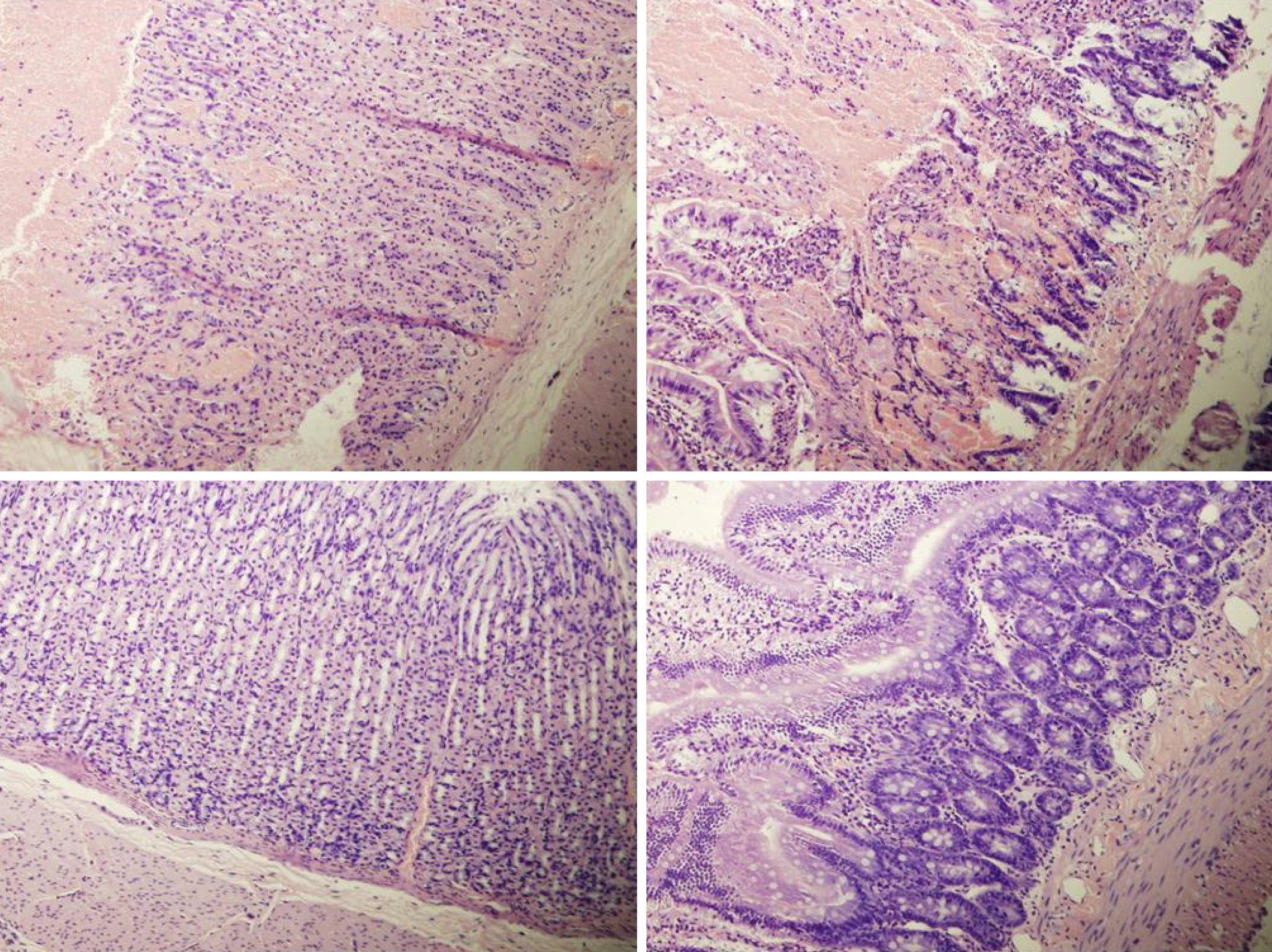
Figure 10 Histological presentation in rats with suprahepatic ligation of the inferior vena cava.
Assessment (stained with hematoxylin-eosin, magnification x 10, scare bar: 50 µm) was at 48 h post-ligation, in controls (upper) and BPC 157-treated rats (lower), stomach (left upper, control; lower, BPC 157), duodenum (right upper, control; lower, BPC 157). Note, substantial congestion and dilatation of mucosal and submucosal capillaries, submucosal edema, ischemic changes, such as architectural distortion and foci of hemorrhage with fibrin deposition in controls (upper), which were markedly counteracted in BPC 157-treated rats (lower).
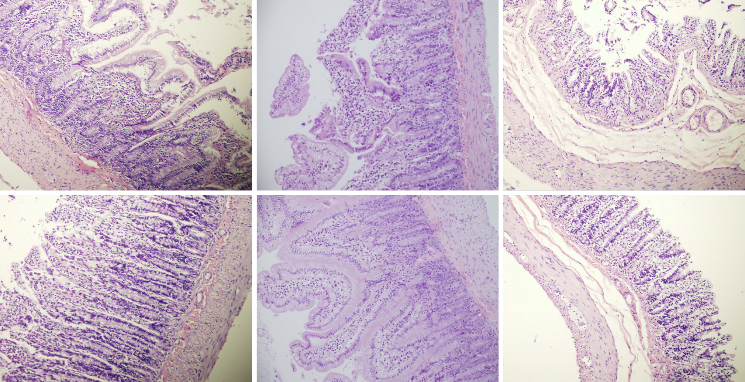
Figure 11 Histological presentation in rats with suprahepatic ligation of the inferior vena cava.
Assessment (stained with hematoxylin-eosin, magnification x 10, scare bar: 50 µm) waste 48 h post-ligation, in controls (upper) and BPC 157-treated rats (lower), jejunum (left upper, control; lower, BPC 157), ileum (middle upper, control; lower, BPC 157), cecum (right upper, control; lower, BPC 157). Note, substantial capillary congestion with mild ischemic changes, loss of crypts with foci of haemorrhage, edema of the lamina propria and mild lymphocytic infiltration in controls (upper), which were markedly counteracted in BPC 157-treated rats (lower).
- Citation: Gojkovic S, Krezic I, Vrdoljak B, Malekinusic D, Barisic I, Petrovic A, Horvat Pavlov K, Kolovrat M, Duzel A, Knezevic M, Kasnik Kovac K, Drmic D, Batelja Vuletic L, Kokot A, Boban Blagaic A, Seiwerth S, Sikiric P. Pentadecapeptide BPC 157 resolves suprahepatic occlusion of the inferior caval vein, Budd-Chiari syndrome model in rats. World J Gastrointest Pathophysiol 2020; 11(1): 1-19
- URL: https://www.wjgnet.com/2150-5330/full/v11/i1/1.htm
- DOI: https://dx.doi.org/10.4291/wjgp.v11.i1.1


