Copyright
©2010 Baishideng Publishing Group Co.
World J Gastrointest Pathophysiol. Oct 15, 2010; 1(4): 129-136
Published online Oct 15, 2010. doi: 10.4291/wjgp.v1.i4.129
Published online Oct 15, 2010. doi: 10.4291/wjgp.v1.i4.129
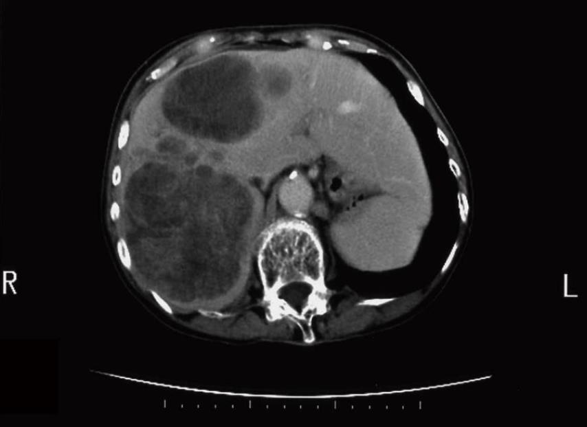
Figure 1 Contrast-enhanced computed tomography image of liver metastasis of protein induced by vitamin K absence or antagonist II-producing gastric cancer.
Multiple low density nodules, which are poorly enhanced, are observed in the liver.
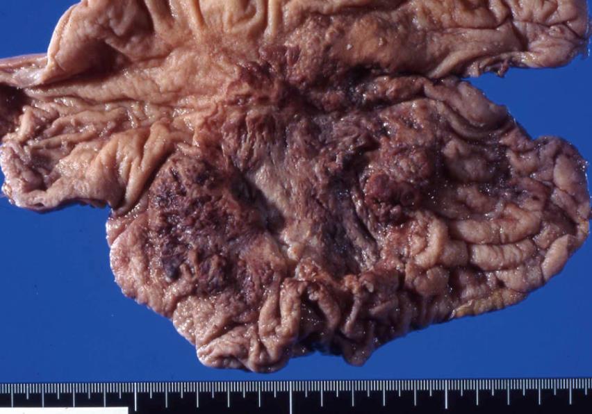
Figure 2 Gross appearance of a case of protein induced by vitamin K absence or antagonist II-producing gastric cancer.
A large Borrmann type 3 tumor occupies the gastric body.
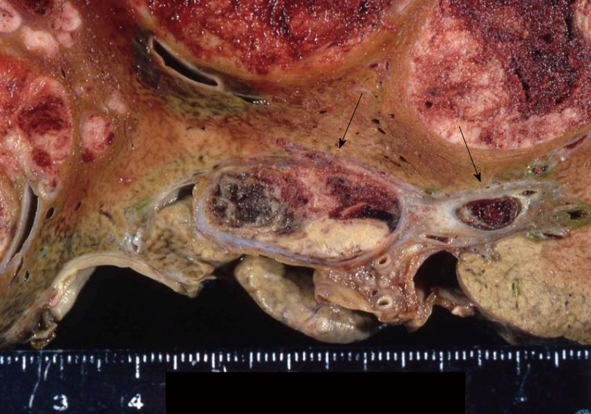
Figure 3 Gross appearance of liver metastasis of protein induced by vitamin K absence or antagonist II-producing gastric cancer.
Multiple nodular tumors and portal vein tumor thrombi (arrows) are observed.
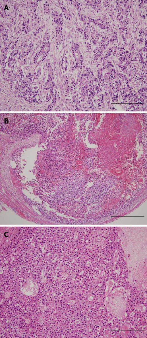
Figure 4 Microscopic appearance of protein induced by vitamin K absence or antagonist II-producing gastric cancer.
A: Gastric tumor with the appearance of a moderately to poorly differentiated adenocarcinoma (HE stain; the scale bar indicates 50 μm); B: Intravenous tumor with a hepatoid pattern (HE stain; the scale bar indicates 100 μm); C: Metastatic liver tumor with a hepatoid pattern (HE stain; the scale bar indicates 50 μm).
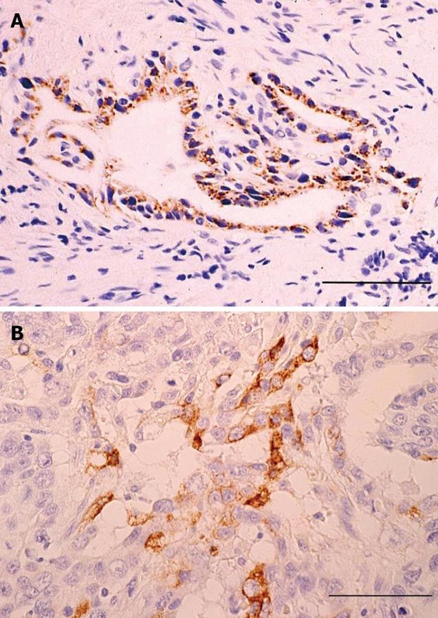
Figure 5 The results of immunohistochemical staining.
A: The tumor cells forming glandular structure are positive for protein induced by vitamin K absence or antagonist II; B: The tumor cells are also positive for alpha-fetoprotein. All scale bars indicate 25 μm.
- Citation: Takahashi Y, Inoue T, Fukusato T. Protein induced by vitamin K absence or antagonist II-producing gastric cancer. World J Gastrointest Pathophysiol 2010; 1(4): 129-136
- URL: https://www.wjgnet.com/2150-5330/full/v1/i4/129.htm
- DOI: https://dx.doi.org/10.4291/wjgp.v1.i4.129


