Copyright
©2010 Baishideng.
World J Gastrointest Pathophysiol. Aug 15, 2010; 1(3): 112-114
Published online Aug 15, 2010. doi: 10.4291/wjgp.v1.i3.112
Published online Aug 15, 2010. doi: 10.4291/wjgp.v1.i3.112
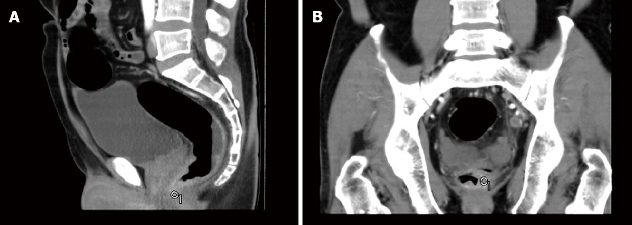
Figure 1 Computed tomography of the abdomen shows a locally, inhomogenously and confoundedly thicken rectal wall (computed tomography value of 35 Hu).
A: Sagittal reconstruction; B: Coronal reconstruction.
Figure 2 Colonoscopic findings.
A, B: Colonoscopic findings indicate a rectal mass encircling the wall of the rectum; C: Follow-up colonoscopy after 3 wk reveals complete regression of the rectal mass.
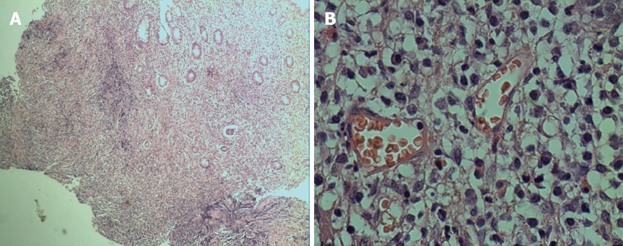
Figure 3 Histological findings from colonoscopic biopsy specimen show diffuse extensive infiltration of a large number of lymphocytes, plasma cells and neutrophil granulocytes (HE staining).
A: × 40; B: × 400.
- Citation: Zhao WT, Liu J, Li YY. Syphilitic proctitis mimicking rectal cancer: A case report. World J Gastrointest Pathophysiol 2010; 1(3): 112-114
- URL: https://www.wjgnet.com/2150-5330/full/v1/i3/112.htm
- DOI: https://dx.doi.org/10.4291/wjgp.v1.i3.112


