Copyright
©2010 Baishideng.
World J Gastrointest Pathophysiol. Aug 15, 2010; 1(3): 106-108
Published online Aug 15, 2010. doi: 10.4291/wjgp.v1.i3.106
Published online Aug 15, 2010. doi: 10.4291/wjgp.v1.i3.106
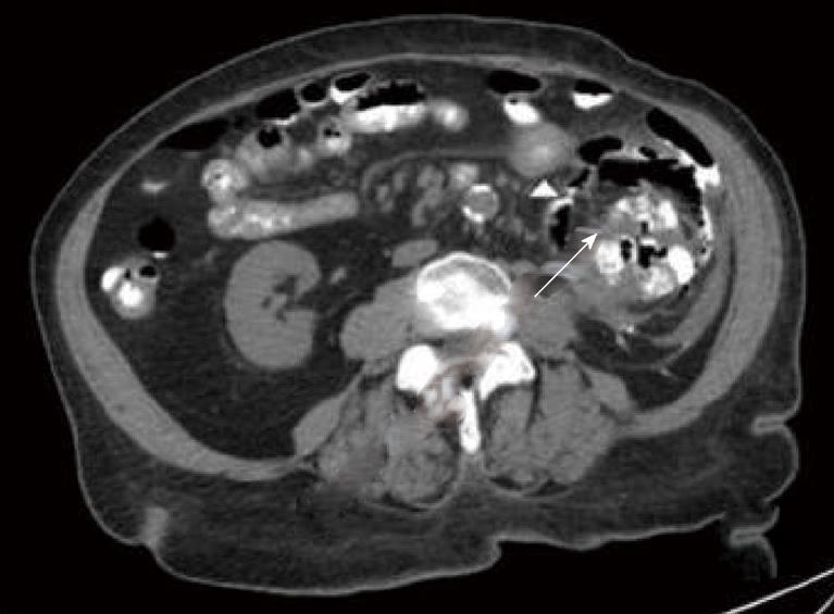
Figure 1 This image illustrates the reflux of the oral contrast into the left kidney via the colo-renal fistula, as well as free air in the renal parenchyma and perinephric space (arrow).
Also noted is thickened small bowel wall adjacent to the left kidney (arrow tip).
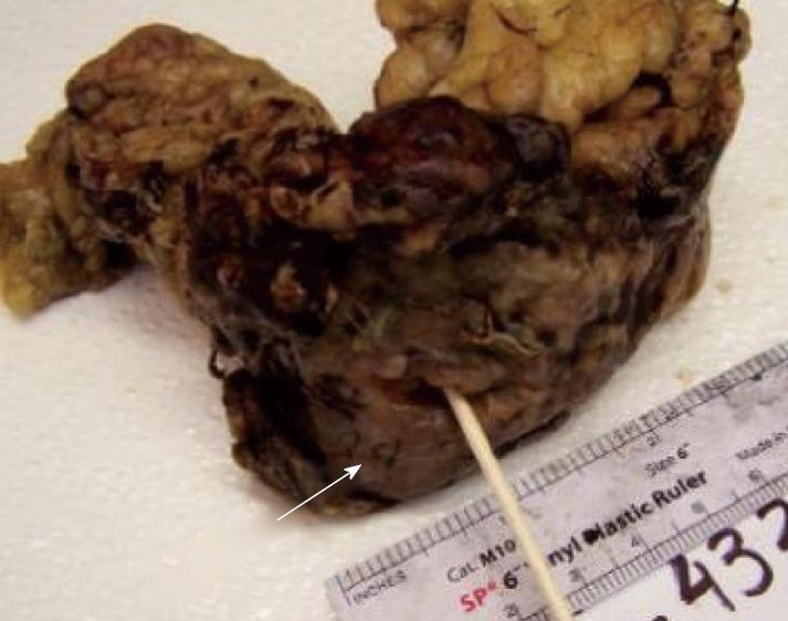
Figure 2 This is a gross specimen of the descending colon that fistulized with the kidney.
There are granular and smoothing changes of the large bowel encompassing the fistula, indicating that there was adhesion and phlegmon prior to fistulization (arrow).
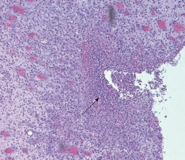
Figure 3 The histopathology represented is a tissue sample from the intestinal fistula.
Note the inflammation on the serosal layer with an accumulation of neutrophils (arrow).
- Citation: Wysocki JD, Joshi V, Eiser JW, Gil N. Colo-renal fistula: An unusual cause of hematochezia. World J Gastrointest Pathophysiol 2010; 1(3): 106-108
- URL: https://www.wjgnet.com/2150-5330/full/v1/i3/106.htm
- DOI: https://dx.doi.org/10.4291/wjgp.v1.i3.106


