Copyright
©2010 Baishideng.
World J Gastrointest Pathophysiol. Jun 15, 2010; 1(2): 38-49
Published online Jun 15, 2010. doi: 10.4291/wjgp.v1.i2.38
Published online Jun 15, 2010. doi: 10.4291/wjgp.v1.i2.38
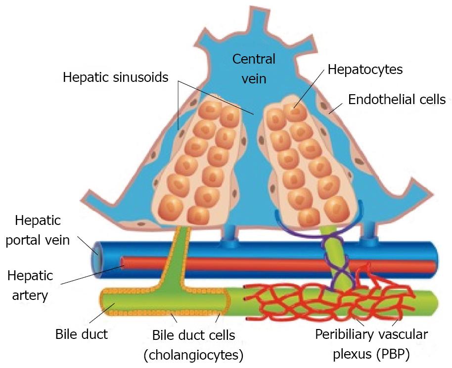
Figure 1 Graphic image of blood flow through the liver and the key cells involved.
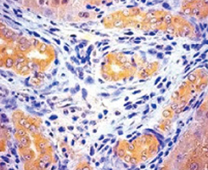
Figure 2 Immunohistochemistry for the H3 histamine receptor in proliferating cholangiocytes (induced by bile duct ligation).
Original magnification × 40.
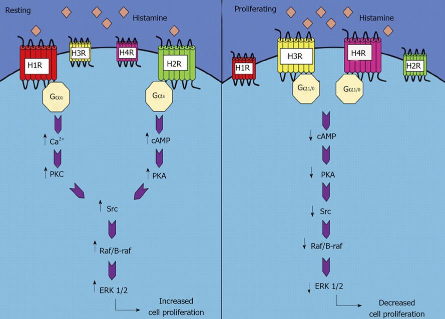
Figure 3 Differential signaling of histamine receptors in resting and proliferating cholangiocytes.
Reprinted with permission from Demorrow S, Francis H, Alpini G, Biogenic Amine Actions on Cholangiocyte Function. Exp Biol Med (Maywood) 2007; 232: 1005-1013.
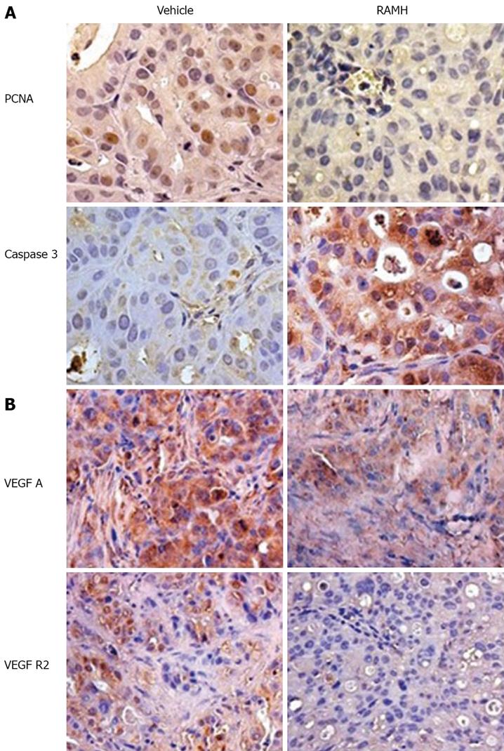
Figure 4 Immunohistochemistry images of tumors excised from vehicle- and RAMH-treated mice (× 40).
A: Proliferating cholangiocytes (PCNA) and apoptotic cholangiocytes (Caspase 3). RAMH induced a decrease in PCNA-positive cholangiocytes coupled with an increase in apoptotic cholangiocytes compared to vehicle-treated mice; B: VEGF-A and VEGF-R2. RAMH treatment caused a decrease in the expression of both VEGF-A and VEGF-R2.
- Citation: Onori P, Gaudio E, Franchitto A, Alpini G, Francis H. Histamine regulation of hyperplastic and neoplastic cell growth in cholangiocytes. World J Gastrointest Pathophysiol 2010; 1(2): 38-49
- URL: https://www.wjgnet.com/2150-5330/full/v1/i2/38.htm
- DOI: https://dx.doi.org/10.4291/wjgp.v1.i2.38


