Copyright
©2010 Baishideng.
World J Gastrointest Pathophysiol. Apr 15, 2010; 1(1): 3-11
Published online Apr 15, 2010. doi: 10.4291/wjgp.v1.i1.3
Published online Apr 15, 2010. doi: 10.4291/wjgp.v1.i1.3
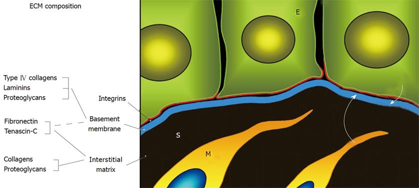
Figure 1 ECM composition and organization of the epithelial-stromal interface in the intestinal mucosa.
The basement membrane (in blue) is located at the interface between the epithelium (E) and the stroma (S). It contains exclusive components such as the type IV collagens and laminins as well as non-exclusive components, such as the fibronectins and tenascins that are also found in the interstitial matrix. Epithelial cells and subepithelial myofibroblasts (M) produce specific basement membrane components. Epithelial cells possess specific receptors for BM components such as the integrins (in red) that are responsible for the mediation of cell-matrix interactions. Adapted from references [4,5].
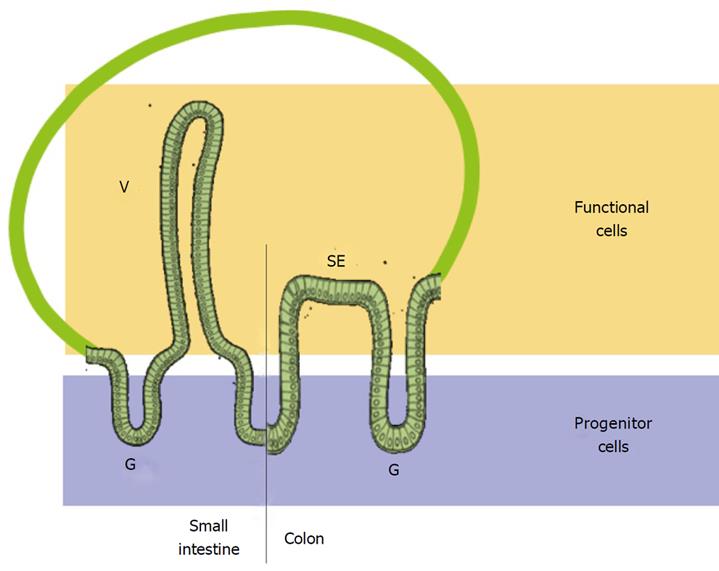
Figure 2 Organization of the renewal units of the human adult small intestine and colon.
In the small intestine, the crypt-villus axis consists of stem and progenitor cells, located in the lower and middle third of the gland (G), respectively, with terminal differentiation occurring in the upper third so that all villus (V) cells are mature and functional. In the colon, the stem cells are located at the bottom of the glands (G) while the proliferative cell compartment extends to the middle. Cells in the upper half of the glands and the surface epithelium (SE) are non-proliferative and functional. Adapted from reference [5].
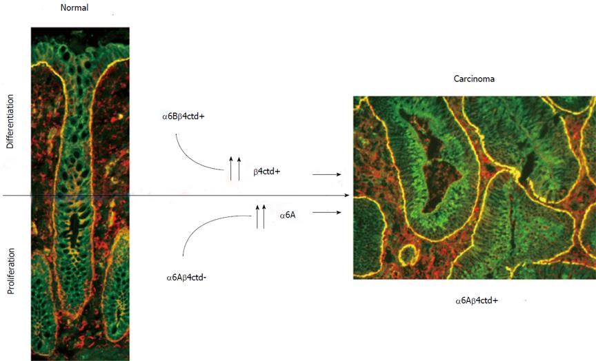
Figure 3 Colorectal cancer cells express a form of α6β4 that is not found in normal colonic cells.
In the normal colon, progenitor cells express the α6Aβ4ctd- form which is pro-proliferative but not functional for adhesion while differentiated cells express the α6Bβ4ctd+ form which is anti-proliferative and functional for adhesion. In colorectal carcinoma, the predominant form of α6β4 is α6Aβ4ctd+, which is pro-proliferative and functional for adhesion.
- Citation: Beaulieu JF. Integrin α6β4 in colorectal cancer. World J Gastrointest Pathophysiol 2010; 1(1): 3-11
- URL: https://www.wjgnet.com/2150-5330/full/v1/i1/3.htm
- DOI: https://dx.doi.org/10.4291/wjgp.v1.i1.3


