Published online Oct 28, 2016. doi: 10.4329/wjr.v8.i10.819
Peer-review started: March 31, 2016
First decision: May 17, 2016
Revised: May 31, 2016
Accepted: August 27, 2016
Article in press: August 29, 2016
Published online: October 28, 2016
Processing time: 211 Days and 0.1 Hours
Blunt diaphragmatic lesions (BDL) are uncommon in trauma patients, but they should be promptly recognized as a delayed diagnosis increases morbidity and mortality. It is well known that BDL are often overlooked at initial imaging, mainly because of distracting injuries to other organs. Sonography may directly depict BDL only in a minor number of cases. Chest X-ray has low sensitivity in detecting BDL and lesions can be reliably suspected only in case of intra-thoracic herniation of abdominal viscera. Thanks to its wide availability, time-effectiveness and spatial resolution, multi-detector computed tomography (CT) is the imaging modality of choice for diagnosing BDL; several direct and indirect CT signs are associated with BDL. Given its high tissue contrast resolution, magnetic resonance imaging can accurately depict BDL, but its use in an emergency setting is limited because of longer acquisition times and need for patient’s collaboration.
Core tip: Blunt diaphragmatic lesions (BDL) are uncommon, but they should be promptly recognized as a delayed diagnosis increases morbidity and mortality. We herein discuss multi-modality imaging findings in BDL and possible pitfalls in order to help the radiologist in this sometimes-difficult diagnosis.
- Citation: Bonatti M, Lombardo F, Vezzali N, Zamboni GA, Bonatti G. Blunt diaphragmatic lesions: Imaging findings and pitfalls. World J Radiol 2016; 8(10): 819-828
- URL: https://www.wjgnet.com/1949-8470/full/v8/i10/819.htm
- DOI: https://dx.doi.org/10.4329/wjr.v8.i10.819
Blunt diaphragmatic lesions (BDL) represent a relatively uncommon event, with estimated prevalence of 0.2%-7% in patients admitted to an Emergency Department (ED) because of blunt trauma[1-3]. Although BDL are rarely life-threatening by themselves in the acute phase, patients suffering from BDL show high mortality rates, ranging from 5% to 50%, because of associated abdominal, thoracic and brain injuries[4-6]. Every diaphragmatic lesion should undergo surgical reparation as soon as possible in order to prevent further complications, like intestinal obstruction or respiratory distress, and to improve patient outcome[7]. Despite the need for an early diagnosis, BDL remain undiagnosed at initial imaging in 7%-66% of the cases[8-10], mainly because of the presence of distracting injuries. The aim of our work is to review imaging findings and possible pitfalls in patients with BDL in order to improve correct diagnosis rates.
The diaphragm represents the anatomical landmark between thoracic and abdominal cavities and serves as the main muscle for respiration. Bilaterally, the diaphragm is composed by three main muscular groups, originating from the lumbar vertebrae, from the inferior border of the ribs and from the sternum, that converge into a thin strong central tendinous sheet. Bilaterally, in the sites where these muscular groups converge, the so-called lumbo-costal triangles and sterno-costal triangles, muscular fibers are replaced by thin areolar tissue, which constitutes a physiological weakness point. Moreover, the posterolateral area of each hemidiaphragm represents a weakness point also being the site where, during the 8th gestational week, the pleuroperitoneal membranes join with the septum trasversum to close the pleuroperitoneal hiatus.
BDL are usually the consequence of high-energy blunt abdominal traumas that determine a sudden increase in abdominal pressure[11], and motor-vehicle collisions represent the most common mechanism of injury. Abdominal pressure is transmitted to the diaphragm that may crack in its physiological weakness points. Less common traumatic mechanisms are represented by high-energy lateral impacts that cause chest wall distortion and subsequent diaphragm shearing, and by direct lacerations from fractured ribs.
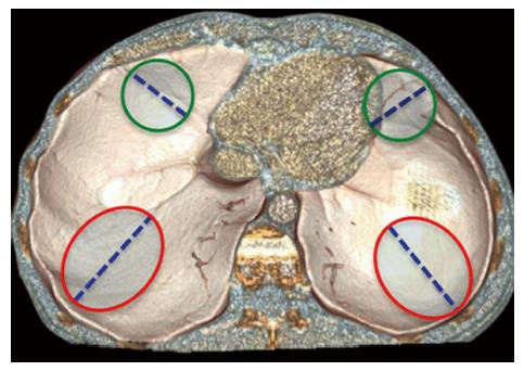
In the majority of cases BDL show a radial course, starting from the postero-lateral areas of the diaphragm and extending towards the central ones (Figure 1) for at least 10 cm[12]. The breakage flaps usually remain close to each other during the early post-traumatic stages, but afterwards they tend to move away because of the difference between the positive intra-abdominal pressure and the negative pleural space one that causes intra-thoracic visceral herniation.
Left BDL are significantly more frequent than right ones (2:1-7:1 ratios), probably because of the protective effect of the liver on the right hemidiaphragm and of a relative weakness of the left hemidiaphragm itself. This difference might also be the consequence of a bias in patient selection: Indeed, right BDL remain undiagnosed at initial imaging with a significantly higher prevalence than left ones[13].
BDL are often asymptomatic in their acute phase, but rarely occur as isolated injuries, as other life-threatening lesions are associated in 50%-100% of cases. Splenic, hepatic and renal injuries are the most commonly associated abdominal lesions, whereas hemothorax, pneumothorax and rib fractures are the most frequent thoracic ones.
Intra-thoracic herniation of abdominal viscera represents a near-invariable consequence of BDL, which may manifest from few seconds to many months after trauma. Intra-thoracic viscera herniation also represents the premise for further complications like intestinal incarceration, strangulation and occlusion, but also respiratory insufficiency.
Being a clinical diagnosis of BDL virtually impossible, imaging plays a central role for this goal. However, it is well known that BDL often remains unrecognized at initial imaging, mainly because of the presence of distracting injuries, and may become manifest only later because of complications[5,14]. Delayed presentation is associated with increased morbidity and mortality[15,16].
Treatment of BDL implicates laparotomy in the majority of the cases, whereas laparoscopy and thoracotomy are less frequently performed[6]. Laparotomy is preferred in hemodynamically unstable patients and in patients with associated intra-abdominal injuries[11]. First of all the surgeon must carefully examine both hemidiaphragms and, in case of intra-thoracic viscera herniation, he must reposition the herniated viscera into the abdominal cavity. The diaphragmatic rupture can then be repaired by means of interrupted or continuous non-absorbable sutures and, finally, a chest tube should be placed in the affected pleural cavity. In case of large diaphragmatic rupture a prosthetic patch can be used for closing the gap.
Chest X-ray (CXR) is performed directly in the shock room in the majority of patients admitted to the ED because of blunt trauma[17]. Given the acute setting and the general conditions of the patients, CXR is usually performed by means of a single antero-posterior projection with the patient lying supine.
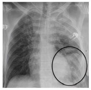
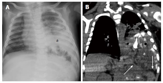
CXR has an unsatisfactory accuracy in detecting BDL; indeed, it may appear normal or it may show only nonspecific changes in 20%-50% of the patients affected by diaphragmatic rupture[18-21]. The only direct sign of BDL at chest radiogram is represented by the visualization of herniated bowel loops into the thoracic cavity (Figure 2). Bowel loops herniation may be appreciated directly or indirectly, because of nasogastric tube apex dislocation.
Some indirect signs might also be present, suggesting the possibility of BDL, but their sensitivity is extremely variable and their specificity is usually unsatisfactory. For example, hemidiaphragm elevation is strongly associated with BDL and previously published studies reported diagnostic accuracies of about 60%[9,22], but its specificity is extremely low, being often present in patients suffering from thoracic trauma. Other low-specificity signs associated with BDL are costophrenic sulcus obliteration and distorted diaphragmatic profile. In any case, the comparison with pre-trauma scans, whenever available, may increase the diagnostic performance by highlighting eventual post-traumatic changes.
Sonography, usually in the form of fast scan, represents the first line imaging modality performed in the majority of blunt trauma patients admitted to the ED. At sonography the diaphragm can be recognized as a curved hyperechoic structure, with a homogeneous thickness, dividing the abdominal cavity from the thoracic one.
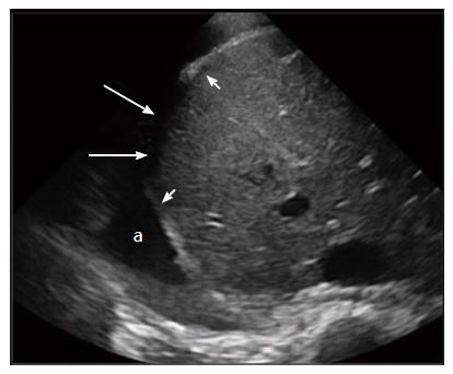
If a good thoracic acoustic window is present (i.e., in case of large pleural effusions), in patients with large diaphragmatic ruptures ultrasound may directly depict the lesion as a focal interruption of the hyperechoic diaphragmatic line (Figure 3). Floating diaphragm edges and intra-thoracic visceral herniation may also be observed[23-25]. On the other hand, small diaphragmatic lesions can be hardly identified and the interpretation of diaphragmatic thickenings remains extremely debated.
Besides the direct visualization of the rupture, some indirect sonographic signs have also been associated with BDL. The impossibility of visualizing one or more hypochondriac organs or the heart without any particular technical factor able to explain it (the so-called “Rip’s absent organ sign”[24]) in a patient admitted to the ED because of a severe blunt abdominal trauma, as well as the hypomobility of one hemidiaphragm in comparison to the other, should raise the suspicion of BDL. In these cases, further examinations, and in particular the performance of a contrast-enhanced multidetector computed tomography (MDCT) scan, are mandatory.
MDCT has nowadays become the imaging modality of choice in trauma patients and also for diagnosing BDL. Indeed, thanks to the technological developments that lead to the introduction of multidetector scanners in the clinical practice, CT offers high accuracy (61%-87% sensitivity and 72%-100% specificity) in diagnosing BDL[26]. CT scans should be acquired with the patient lying supine on the table, with the hands over its head whenever possible; otherwise, the arms should be bent over a pillow on his abdomen in order to reduce beam hardening artifacts. The acquisition should be as fast as possible in order to prevent motion artifacts[27]. Contrast material administration increases the conspicuity between different anatomical structures and, therefore, it is useful in order to better delimitate the diaphragm. Moreover, the evaluation of multiplanar reconstructions on sagittal and coronal planes increases CT accuracy in BDL detection[28,29].
CT often allows to directly depict diaphragmatic lesions as segmental defects; other direct signs of BDI are diaphragm non-visualization, dangling diaphragm sign and diaphragm thickening. Many different indirect signs have also been associated with BDL, i.e., intra-thoracic viscera herniation, dependent viscera sign, collar sign and hump and band sign.
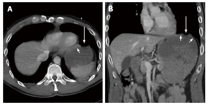
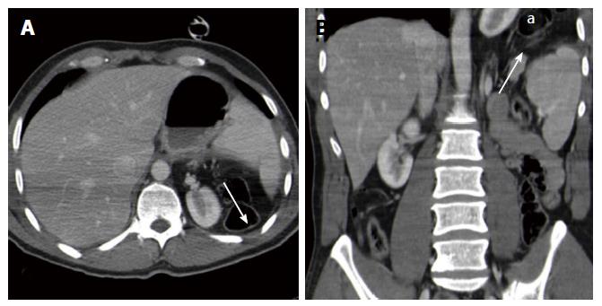
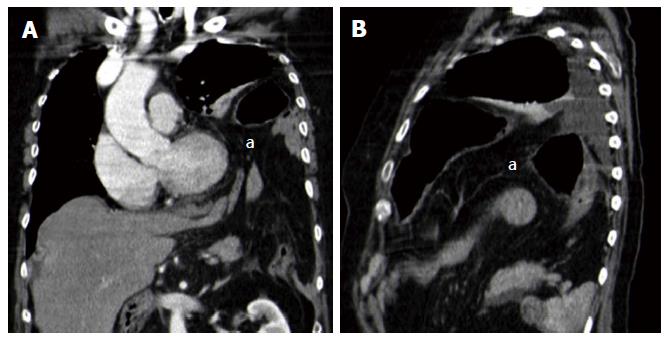
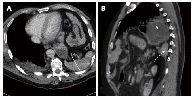
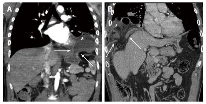
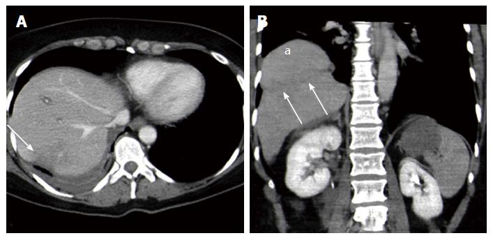
A segmental diaphragmatic defect (Figure 4) represents the most commonly encountered sign of BDL, with sensitivity values up to 95.7% in the most recent Literature[26,30]. The defect can be better appreciated on multiplanar reconstructions performed perpendicular to the tear than on axial images[31]. In the majority of the cases, the defect is delimited by slightly thickened diaphragmatic edges, as a consequence of edema, intramuscular hematoma and muscle retraction. Attention must be paid to physiological diaphragmatic defects, which may be observed in up to 11% of healthy population, with a prevalence that increases with age[32]: In these cases, no edge thickening is observed. In case of large diaphragmatic lesions, partial or complete diaphragm non-visualization (Figure 5) represents a relatively frequent event and it is almost always associated with intra-thoracic viscera herniation. The retracted diaphragm may be sometimes recognized at its bone insertion. The sensitivity of this sign is not as high as that of a simple diaphragmatic defect, but its specificity approaches 100%. Intra-thoracic viscera herniation (Figure 6) represents the direct consequence of the difference between intra-abdominal and intra-thoracic pressure, which brings the abdominal organs to herniate in the thoracic cavity if a large-enough diaphragmatic lesion is present. Viscera herniation is more common on the left than on the right side, because of the lack of the protective effect of the liver. Herniation probability increases parallel to tear extension. Intrapericardial herniation represents a rare but potentially fatal event[19].
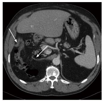
Diaphragm thickening (Figure 7) is usually present in association with other signs of diaphragm rupture, but it may also be the only finding of small BDL. In high-energy blunt traumas, diaphragm thickness should be always accurately compared with the contralateral side in order to rule out a lesion. Diaphragm thickening may be the consequence of muscular edema or hematoma, but it may also be the indicator of a still-closed diaphragmatic tear; therefore, this finding should be always highlighted in the radiological report and, in case of patients without any other indication for surgical exploration, it must be strictly followed-up by means of CT. Anyway, some variability in diaphragmatic thickness may be physiological[12].
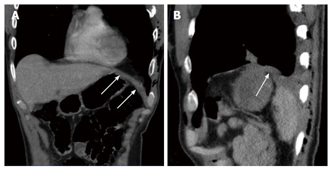
The dangling diaphragm sign (Figure 8) was first described by Desser et al[33] in 2010 as the visualization of the free edges of the torn diaphragm as comma-shaped structures, which curl inward, toward the center of the abdomen. It is usually associated with a segmental diaphragmatic defect and diaphragm thickening. The dangling diaphragm sign may be observed in only about 50% of the patients with BDL, but its specificity reaches 98%[26,30,33].
The dependent viscera sign[34] (Figure 9) (also referred to as sinus cutoff sign[35]) was first described by Bergin et al[34] in 2001 and is represented by an anomalous contact between abdominal viscera and posterior chest wall, without the physiological lung parenchyma interposition. It is the consequence of the loss of the diaphragmatic support to the abdominal organs that, therefore, in the supine position tend to lie dorsally. It was particularly useful for BDL detection in the pre-multiplanar reconstructions era.
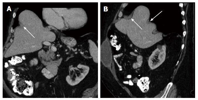
The collar sign (also referred to as hourglass sign) (Figure 10) is represented by a “waist-like” constriction of the herniated viscera at the point of diaphragmatic discontinuity and is typically observed in case of left diaphragmatic lesions with gastric herniation. The hump and band sign (Figure 11) is similar to the collar sign and refers to the shape of the herniated liver in case of right BDL. The hump shape is due to the squeezing of the herniated liver above the diaphragmatic defect, whereas the hypoattenuating band is thought to be the consequence of local liver parenchyma hypoperfusion because of diaphragmatic compression. It is best appreciated on sagittal and coronal reconstructions.
The radiologist must pay particular attention to the above-mentioned signs in patients with parenchymal organs lesions, rib fractures, pleural effusion, hemothorax or hemoperitoneum.
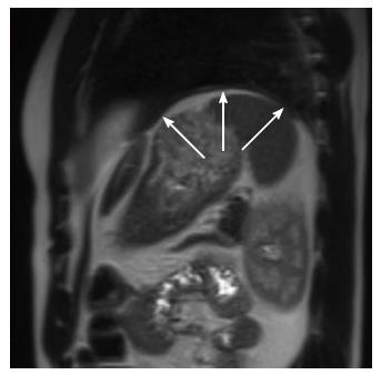
Magnetic resonance imaging (MRI) warrants higher tissue contrast resolution than CT and enables to clearly depict the diaphragm (Figure 12), but its use in an emergency setting is limited by the longer acquisition times and by the need for patient collaboration. Moreover, the original advantage of MRI in comparison with CT in diagnosing BDL, i.e., its multiplanarity, has been overwhelmed by the development of multidetector CT scanners. Therefore, nowadays MRI should be considered a second level investigation reserved to hemodynamically stable patients with inconclusive findings at CT[36].
In order to better evaluate the hemidiaphragms, MRI sequences should be acquired using cardiac and respiratory gating. The examination can be simply based on spin-echo T1-weighted sequences, acquired on the axial, sagittal and coronal planes, in which the normal diaphragm appears as a continuous hypointense band surrounded by hyperintense abdominal and subpleural fat[37]. In this setting, diaphragmatic discontinuities as well as the other findings described for CT may be accurately recognized[38,39]. Gradient-echo T1-weighted sequences represent an alternative to SE ones because of faster acquisition times, but chemical shift artifacts may limit their value.
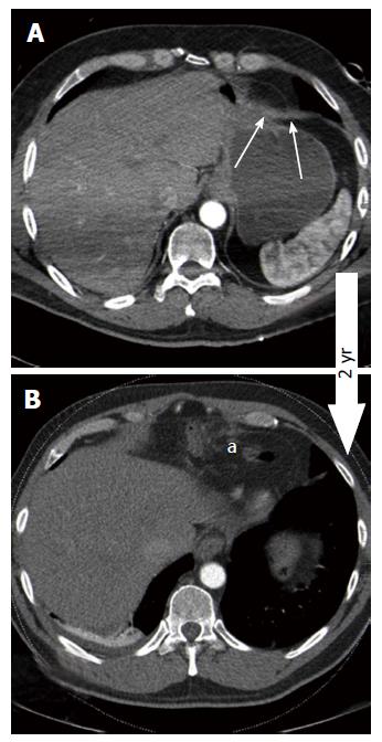
Slight alterations, like slight diaphragmatic thickenings (Figure 13) or small diaphragmatic defects, may often remain undiagnosed at initial imaging, especially if major distracting injuries are present, and may manifest only later because of complications. In the matter of facts, the presence of severe thoraco-abdominal injuries should point the radiologist’s attention to the diaphragm.
BDL diagnosis may be more difficult in patients receiving mechanical ventilation, in particular if positive pressures are adopted[40]. Indeed, mechanical ventilation reduces the difference between intra-abdominal and intra-thoracic pressure, preventing abdominal viscera herniation (Figure 14) and reducing lesion conspicuity.
Eventration (Figure 15) may involve an entire hemidiaphragm or only a part of it. In both cases muscle continuity must be clearly recognizable and no abnormal diaphragmatic thickenings must be observed; on the other hand, in case of eventration the involved diaphragm is usually uniformly thinned.
Congenital hernias (Figure 16) are usually located in the same areas where traumatic lesions occur and, therefore, the differential diagnosis may be particularly challenging. However, congenital hernias are usually not associated with diaphragm thickening.
In the era of multidetector CT and of high quality multiplanar reconstructions, the diagnosis of BDL should be no more overlooked by radiologists working in an emergency setting. Appropriate knowledge of the common and less common signs of BDL and careful evaluation of the diaphragm in patients with blunt trauma should enable a timely and accurate diagnosis.
Manuscript source: Invited manuscript
Specialty type: Radiology, nuclear medicine and medical imaging
Country of origin: Italy
Peer-review report classification
Grade A (Excellent): 0
Grade B (Very good): B, B
Grade C (Good): 0
Grade D (Fair): 0
Grade E (Poor): 0
P- Reviewer: Lovric Z, Slomiany BL S- Editor: Ji FF L- Editor: A E- Editor: Li D
| 1. | Rodriguez-Morales G, Rodriguez A, Shatney CH. Acute rupture of the diaphragm in blunt trauma: analysis of 60 patients. J Trauma. 1986;26:438-444. [RCA] [PubMed] [DOI] [Full Text] [Cited by in Crossref: 126] [Cited by in RCA: 100] [Article Influence: 2.6] [Reference Citation Analysis (0)] |
| 2. | Sliker CW. Imaging of diaphragm injuries. Radiol Clin North Am. 2006;44:199-211, vii. [RCA] [PubMed] [DOI] [Full Text] [Cited by in Crossref: 80] [Cited by in RCA: 65] [Article Influence: 3.4] [Reference Citation Analysis (0)] |
| 3. | Hsee L, Wigg L, Civil I. Diagnosis of blunt traumatic ruptured diaphragm: is it still a difficult problem? ANZ J Surg. 2010;80:166-168. [RCA] [PubMed] [DOI] [Full Text] [Cited by in Crossref: 8] [Cited by in RCA: 9] [Article Influence: 0.6] [Reference Citation Analysis (0)] |
| 4. | Haciibrahimoglu G, Solak O, Olcmen A, Bedirhan MA, Solmazer N, Gurses A. Management of traumatic diaphragmatic rupture. Surg Today. 2004;34:111-114. [RCA] [PubMed] [DOI] [Full Text] [Cited by in Crossref: 53] [Cited by in RCA: 51] [Article Influence: 2.4] [Reference Citation Analysis (0)] |
| 5. | Mihos P, Potaris K, Gakidis J, Paraskevopoulos J, Varvatsoulis P, Gougoutas B, Papadakis G, Lapidakis E. Traumatic rupture of the diaphragm: experience with 65 patients. Injury. 2003;34:169-172. [RCA] [PubMed] [DOI] [Full Text] [Cited by in Crossref: 126] [Cited by in RCA: 110] [Article Influence: 5.0] [Reference Citation Analysis (0)] |
| 6. | Chughtai T, Ali S, Sharkey P, Lins M, Rizoli S. Update on managing diaphragmatic rupture in blunt trauma: a review of 208 consecutive cases. Can J Surg. 2009;52:177-181. [PubMed] |
| 7. | Iochum S, Ludig T, Walter F, Sebbag H, Grosdidier G, Blum AG. Imaging of diaphragmatic injury: a diagnostic challenge? Radiographics. 2002;22 Spec No:S103-S116; discussion S116-S118. [RCA] [PubMed] [DOI] [Full Text] [Cited by in Crossref: 160] [Cited by in RCA: 125] [Article Influence: 5.4] [Reference Citation Analysis (0)] |
| 8. | Desir A, Ghaye B. CT of blunt diaphragmatic rupture. Radiographics. 2012;32:477-498. [RCA] [PubMed] [DOI] [Full Text] [Cited by in Crossref: 84] [Cited by in RCA: 95] [Article Influence: 7.3] [Reference Citation Analysis (0)] |
| 9. | Guth AA, Pachter HL, Kim U. Pitfalls in the diagnosis of blunt diaphragmatic injury. Am J Surg. 1995;170:5-9. [RCA] [PubMed] [DOI] [Full Text] [Cited by in Crossref: 99] [Cited by in RCA: 80] [Article Influence: 2.7] [Reference Citation Analysis (0)] |
| 10. | Murray JG, Caoili E, Gruden JF, Evans SJ, Halvorsen RA, Mackersie RC. Acute rupture of the diaphragm due to blunt trauma: diagnostic sensitivity and specificity of CT. AJR Am J Roentgenol. 1996;166:1035-1039. [RCA] [PubMed] [DOI] [Full Text] [Cited by in Crossref: 133] [Cited by in RCA: 101] [Article Influence: 3.5] [Reference Citation Analysis (0)] |
| 11. | Tan KK, Yan ZY, Vijayan A, Chiu MT. Management of diaphragmatic rupture from blunt trauma. Singapore Med J. 2009;50:1150-1153. [PubMed] |
| 12. | Bocchini G, Guida F, Sica G, Codella U, Scaglione M. Diaphragmatic injuries after blunt trauma: are they still a challenge? Reviewing CT findings and integrated imaging. Emerg Radiol. 2012;19:225-235. [RCA] [PubMed] [DOI] [Full Text] [Cited by in Crossref: 31] [Cited by in RCA: 27] [Article Influence: 2.1] [Reference Citation Analysis (0)] |
| 13. | Walchalk LR, Stanfield SC. Delayed presentation of traumatic diaphragmatic rupture. J Emerg Med. 2010;39:21-24. [RCA] [PubMed] [DOI] [Full Text] [Cited by in Crossref: 13] [Cited by in RCA: 19] [Article Influence: 1.1] [Reference Citation Analysis (0)] |
| 14. | Kuo IM, Liao CH, Hsin MC, Kang SC, Wang SY, Ooyang CH, Fang JF. Blunt diaphragmatic rupture--a rare but challenging entity in thoracoabdominal trauma. Am J Emerg Med. 2012;30:919-924. [RCA] [PubMed] [DOI] [Full Text] [Cited by in Crossref: 19] [Cited by in RCA: 19] [Article Influence: 1.5] [Reference Citation Analysis (0)] |
| 15. | Ganie FA, Lone H, Lone GN, Wani ML, Ganie SA, Wani NU, Gani M. Delayed presentation of traumatic diaphragmatic hernia: a diagnosis of suspicion with increased morbidity and mortality. Trauma Mon. 2013;18:12-16. [RCA] [PubMed] [DOI] [Full Text] [Full Text (PDF)] [Cited by in Crossref: 19] [Cited by in RCA: 25] [Article Influence: 2.1] [Reference Citation Analysis (0)] |
| 16. | Brasel KJ, Borgstrom DC, Meyer P, Weigelt JA. Predictors of outcome in blunt diaphragm rupture. J Trauma. 1996;41:484-487. [RCA] [PubMed] [DOI] [Full Text] [Cited by in Crossref: 23] [Cited by in RCA: 19] [Article Influence: 0.7] [Reference Citation Analysis (0)] |
| 17. | Aukema TS, Beenen LF, Hietbrink F, Leenen LP. Initial assessment of chest X-ray in thoracic trauma patients: Awareness of specific injuries. World J Radiol. 2012;4:48-52. [RCA] [PubMed] [DOI] [Full Text] [Full Text (PDF)] [Cited by in CrossRef: 15] [Cited by in RCA: 15] [Article Influence: 1.2] [Reference Citation Analysis (0)] |
| 18. | Shackleton KL, Stewart ET, Taylor AJ. Traumatic diaphragmatic injuries: spectrum of radiographic findings. Radiographics. 1998;18:49-59. [RCA] [PubMed] [DOI] [Full Text] [Cited by in Crossref: 71] [Cited by in RCA: 49] [Article Influence: 1.8] [Reference Citation Analysis (0)] |
| 19. | Mirvis SE, Shanmuganagthan K. Imaging hemidiaphragmatic injury. Eur Radiol. 2007;17:1411-1421. [RCA] [PubMed] [DOI] [Full Text] [Cited by in Crossref: 45] [Cited by in RCA: 40] [Article Influence: 2.2] [Reference Citation Analysis (0)] |
| 20. | Sangster G, Ventura VP, Carbo A, Gates T, Garayburu J, D’Agostino H. Diaphragmatic rupture: a frequently missed injury in blunt thoracoabdominal trauma patients. Emerg Radiol. 2007;13:225-230. [RCA] [PubMed] [DOI] [Full Text] [Cited by in Crossref: 38] [Cited by in RCA: 33] [Article Influence: 1.7] [Reference Citation Analysis (0)] |
| 21. | Langdorf MI, Medak AJ, Hendey GW, Nishijima DK, Mower WR, Raja AS, Baumann BM, Anglin DR, Anderson CL, Lotfipour S. Prevalence and Clinical Import of Thoracic Injury Identified by Chest Computed Tomography but Not Chest Radiography in Blunt Trauma: Multicenter Prospective Cohort Study. Ann Emerg Med. 2015;66:589-600. [RCA] [PubMed] [DOI] [Full Text] [Cited by in Crossref: 56] [Cited by in RCA: 70] [Article Influence: 7.0] [Reference Citation Analysis (0)] |
| 22. | Gelman R, Mirvis SE, Gens D. Diaphragmatic rupture due to blunt trauma: sensitivity of plain chest radiographs. AJR Am J Roentgenol. 1991;156:51-57. [RCA] [PubMed] [DOI] [Full Text] [Cited by in Crossref: 195] [Cited by in RCA: 145] [Article Influence: 4.3] [Reference Citation Analysis (0)] |
| 23. | Blaivas M, Brannam L, Hawkins M, Lyon M, Sriram K. Bedside emergency ultrasonographic diagnosis of diaphragmatic rupture in blunt abdominal trauma. Am J Emerg Med. 2004;22:601-604. [RCA] [PubMed] [DOI] [Full Text] [Cited by in Crossref: 62] [Cited by in RCA: 55] [Article Influence: 2.8] [Reference Citation Analysis (0)] |
| 24. | Gangahar R, Doshi D. FAST scan in the diagnosis of acute diaphragmatic rupture. Am J Emerg Med. 2010;28:387.e1-387.e3. [RCA] [PubMed] [DOI] [Full Text] [Cited by in Crossref: 15] [Cited by in RCA: 9] [Article Influence: 0.6] [Reference Citation Analysis (0)] |
| 25. | Kim HH, Shin YR, Kim KJ, Hwang SS, Ha HK, Byun JY, Choi KH, Shinn KS. Blunt traumatic rupture of the diaphragm: sonographic diagnosis. J Ultrasound Med. 1997;16:593-598. [PubMed] |
| 26. | Panda A, Kumar A, Gamanagatti S, Patil A, Kumar S, Gupta A. Traumatic diaphragmatic injury: a review of CT signs and the difference between blunt and penetrating injury. Diagn Interv Radiol. 2014;20:121-128. [RCA] [PubMed] [DOI] [Full Text] [Cited by in Crossref: 13] [Cited by in RCA: 20] [Article Influence: 2.0] [Reference Citation Analysis (0)] |
| 27. | Liang T, McLaughlin P, Arepalli CD, Louis LJ, Bilawich AM, Mayo J, Nicolaou S. Dual-source CT in blunt trauma patients: elimination of diaphragmatic motion using high-pitch spiral technique. Emerg Radiol. 2016;23:127-132. [RCA] [PubMed] [DOI] [Full Text] [Cited by in Crossref: 6] [Cited by in RCA: 6] [Article Influence: 0.6] [Reference Citation Analysis (0)] |
| 28. | Bhullar IS, Block EF. CT with coronal reconstruction identifies previously missed smaller diaphragmatic injuries after blunt trauma. Am Surg. 2011;77:55-58. [PubMed] |
| 29. | Larici AR, Gotway MB, Litt HI, Reddy GP, Webb WR, Gotway CA, Dawn SK, Marder SR, Storto ML. Helical CT with sagittal and coronal reconstructions: accuracy for detection of diaphragmatic injury. AJR Am J Roentgenol. 2002;179:451-457. [RCA] [PubMed] [DOI] [Full Text] [Cited by in Crossref: 80] [Cited by in RCA: 73] [Article Influence: 3.2] [Reference Citation Analysis (0)] |
| 30. | Hammer MM, Flagg E, Mellnick VM, Cummings KW, Bhalla S, Raptis CA. Computed tomography of blunt and penetrating diaphragmatic injury: sensitivity and inter-observer agreement of CT Signs. Emerg Radiol. 2014;21:143-149. [RCA] [PubMed] [DOI] [Full Text] [Cited by in Crossref: 29] [Cited by in RCA: 26] [Article Influence: 2.2] [Reference Citation Analysis (0)] |
| 31. | Killeen KL, Mirvis SE, Shanmuganathan K. Helical CT of diaphragmatic rupture caused by blunt trauma. AJR Am J Roentgenol. 1999;173:1611-1616. [RCA] [PubMed] [DOI] [Full Text] [Cited by in Crossref: 169] [Cited by in RCA: 139] [Article Influence: 5.3] [Reference Citation Analysis (0)] |
| 32. | Caskey CI, Zerhouni EA, Fishman EK, Rahmouni AD. Aging of the diaphragm: a CT study. Radiology. 1989;171:385-389. [RCA] [PubMed] [DOI] [Full Text] [Cited by in Crossref: 90] [Cited by in RCA: 77] [Article Influence: 2.1] [Reference Citation Analysis (0)] |
| 33. | Desser TS, Edwards B, Hunt S, Rosenberg J, Purtill MA, Jeffrey RB. The dangling diaphragm sign: sensitivity and comparison with existing CT signs of blunt traumatic diaphragmatic rupture. Emerg Radiol. 2010;17:37-44. [RCA] [PubMed] [DOI] [Full Text] [Cited by in Crossref: 29] [Cited by in RCA: 24] [Article Influence: 1.5] [Reference Citation Analysis (0)] |
| 34. | Bergin D, Ennis R, Keogh C, Fenlon HM, Murray JG. The “dependent viscera” sign in CT diagnosis of blunt traumatic diaphragmatic rupture. AJR Am J Roentgenol. 2001;177:1137-1140. [RCA] [PubMed] [DOI] [Full Text] [Cited by in Crossref: 114] [Cited by in RCA: 85] [Article Influence: 3.5] [Reference Citation Analysis (0)] |
| 35. | Kaya SO, Karabulut N, Yuncu G, Sevinc S, Kiroğlu Y. Sinus cut-off sign: a helpful sign in the CT diagnosis of diaphragmatic rupture associated with pleural effusion. Eur J Radiol. 2006;59:253-256. [RCA] [PubMed] [DOI] [Full Text] [Cited by in Crossref: 9] [Cited by in RCA: 9] [Article Influence: 0.5] [Reference Citation Analysis (0)] |
| 36. | Eren S, Kantarci M, Okur A. Imaging of diaphragmatic rupture after trauma. Clin Radiol. 2006;61:467-477. [RCA] [PubMed] [DOI] [Full Text] [Cited by in Crossref: 63] [Cited by in RCA: 46] [Article Influence: 2.4] [Reference Citation Analysis (0)] |
| 37. | Gierada DS, Curtin JJ, Erickson SJ, Prost RW, Strandt JA, Goodman LR. Diaphragmatic motion: fast gradient-recalled-echo MR imaging in healthy subjects. Radiology. 1995;194:879-884. [RCA] [PubMed] [DOI] [Full Text] [Cited by in Crossref: 125] [Cited by in RCA: 110] [Article Influence: 3.7] [Reference Citation Analysis (0)] |
| 38. | Barbiera F, Nicastro N, Finazzo M, Lo Casto A, Runza G, Bartolotta TV, Midiri M. The role of MRI in traumatic rupture of the diaphragm. Our experience in three cases and review of the literature. Radiol Med. 2003;105:188-194. [PubMed] |
| 39. | Shanmuganathan K, Mirvis SE, White CS, Pomerantz SM. MR imaging evaluation of hemidiaphragms in acute blunt trauma: experience with 16 patients. AJR Am J Roentgenol. 1996;167:397-402. [RCA] [PubMed] [DOI] [Full Text] [Cited by in Crossref: 81] [Cited by in RCA: 59] [Article Influence: 2.0] [Reference Citation Analysis (0)] |


