Published online Jun 28, 2015. doi: 10.4329/wjr.v7.i6.131
Peer-review started: January 19, 2015
First decision: February 7, 2015
Revised: March 10, 2015
Accepted: April 10, 2015
Article in press: April 14, 2015
Published online: June 28, 2015
Processing time: 150 Days and 20 Hours
The vasa vasorum of carotid artery plaque is a novel marker of accurately evaluating the vulnerability of carotid artery plaque, which was associated with symptomatic cerebrovascular and cardiovascular disease. The presence of ultrasound contrast agents in carotid artery plaque represents the presence of the vasa vasorum in carotid artery plaque because the ultrasound contrast agents are strict intravascular tracers. Therefore, contrast-enhanced ultrasound (CEUS) is a novel and safe imaging modality for evaluating the vasa vasorum in carotid artery plaque. However, there are some issues that needs to be assessed to embody fully the clinical utility of the vasa vasorum in carotid artery plaque with CEUS.
Core tip: Stroke is a major cause of morbidity and mortality all over the world. At-risk patients is so-called vulnerable patients because they posses a higher likelihood of developing symptomatic stroke compared with those low-risk patients. Vulnerable patients usually have carotid artery vulnerable plaques and vulnerable plaques posses a higher likelihood of rupture to lead to acute stroke. The vasa vasorum of carotid artery plaque has been confirmed as same as vulnerable plaques and contrast-enhanced ultrasonography could detect the vasa vasorum of carotid artery plaque.
- Citation: Song ZZ, Zhang YM. Contrast-enhanced ultrasound imaging of the vasa vasorum of carotid artery plaque. World J Radiol 2015; 7(6): 131-133
- URL: https://www.wjgnet.com/1949-8470/full/v7/i6/131.htm
- DOI: https://dx.doi.org/10.4329/wjr.v7.i6.131
The plaques with a thin fibrous cap covering a large lipid necrotic core, and active inflammation is so-called vulnerable atherosclerotic plaque, which that make the plaque at increased risk of rupture[1]. The vasa vasorum of carotid artery plaque is a novel marker of accurately evaluating the vulnerability of carotid artery plaque[1,2]. The presence of vasa vasorum in carotid artery plaque has been associated with symptomatic cerebrovascular and cardiovascular disease[3,4] because the vasa vasorum in carotid artery plaque is increased risk for rupture, causing intraplaque hemorrhage and subsequent rapid progression to symptomatic disease.
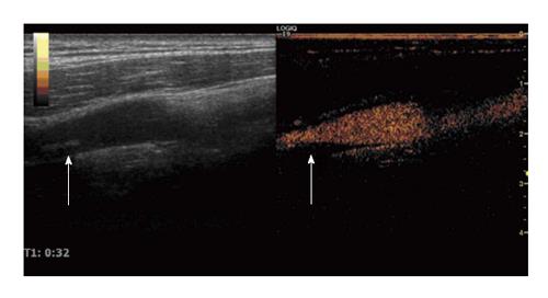
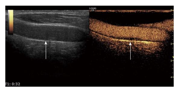
Recently, contrast-enhanced ultrasound (CEUS) has been introduced for identifying the presence of the vasa vasorum in carotid artery plaque[5-9], therefore, CEUS is capable of assessing atherosclerotic carotid lesions at risk for rupture[5,10]. The currently approved and used agents are SonoVue (Bracco SpA, Milan, Italy) in china. Ultrasound contrast agents have been administrated in millions of patients and are safe and side effects are extremely rare[11]. The presence of ultrasound contrast agents in carotid artery plaque represents the presence of the vasa vasorum in carotid artery plaque (Figures 1 and 2) because the ultrasound contrast agents (SonoVue) are strict intravascular tracers, and the appearance of contrast enhancement of CEUS was shown to correlate with the presence and degree of the vasa vasorum in carotid artery plaque which were assessed by histology[12,13].
After implemented in the routine carotid ultrasound scan acquisition protocol, CEUS of carotid artery plaque can be relatively straightforward[8,9]. Firstly, a venous access catheter is placed into median vein of elbow. Secondly, the contrast presets of the ultrasound system are selected, which are available in nearly all currently used vascular ultrasound systems. Carotid CEUS imaging usually uses a linear array transducer that transmits frequencies are between 5 and 10 MHz and a low-level mechanical index condition is used to avoid destruction of the microbubbles[8,9]. Thirdly, high quality CEUS dynamical images of the continuous appearance of carotid artery plaque can be obtained after ultrasound contrast agent followed by a 5 mL saline flush are injected into median vein of elbow according to venous access catheter[8,9]. Usually, the optimal time window for the performance of CEUS after administration of the contrast agent is approximately in 2 min[8,9]. Lastly, the ultrasound contrast signal intensity becomes weaker because the ultrasound contrast agent is eliminated after a few minutes, and administration of the ultrasound contrast agent can be repeated[8,9].
There are too many criterions about assessment of the vasa vasorum in carotid artery plaque with CEUS at the present[4-9,12,13], which includes quantitative criterion, semi-quantitative criterion and qualltative criterion[4-9], therefore, the criterion of assessment of the vasa vasorum in carotid artery plaque with CEUS is inconsistent among all kinds of clinical studies[4-9,12,13]. Thus, the clinical value of the vasa vasorum in carotid artery plaque with CEUS yet remain sure accurately, therefore, the criterion about assessment of the vasa vasorum in carotid artery plaque with CEUS should be assessed in the future studies.
The dosage of ultrasound contrast agent was different among some studies[4-9]. To our experience[8,9], 1.2 mL ultrasound contrast agent followed by a 5 mL saline flush acquire high quality CEUS dynamical images of the continuous appearance of carotid artery plaque, although some studies and authors propose 2.4 mL or 2.0 mL ultrasound contrast agent followed by a 5 mL saline flush[4-6,12,13]. However, the different dosage of ultrasound contrast agent could cause the results according to quantitative criterion, semi-quantitative criterion and qualltative criterion significantly discrepancy, therefore, the dosage of ultrasound contrast agent should be unified according to the quality of CEUS dynamical images and cost-effectiveness.
The clinical studies[4-9,12,13] about the vasa vasorum in carotid artery plaque with CEUS are exciting because the authors think the method could provide early detection and classification of atherosclerotic disease, however, consistency and repetition of the method is poorly established[4-9,12,13]. The lowness of consistency and repetition could cause the method not extensive use, therefore, consistency and repetition of the method needs to be assessed in prospective studies.
It is well known that the vasa vasorum in carotid artery plaque with CEUS were well correlated with histologic specimens obtained after endarterectomy[5-7], however, the diagnosis accuracy of the histologic degree of the vasa vasorum in carotid artery plaque using CEUS remain unclear. Therefore, this issue should be assessed in the future perspective study.
Most of the studies about the vasa vasorum in carotid artery plaque with CEUS are retrospective studies[7-9], therefore, too many issues about the clinical utility of the vasa vasorum in carotid artery plaque with CEUS remain unclear. In addition, most of studies about the vasa vasorum in carotid artery plaque with CEUS are small-scale, which causes the results of these studies unimpressive. Therefore, the large-scale perspective studies should be performed to assess the clinical utility of the vasa vasorum in carotid artery plaque with CEUS.
CEUS is a novel and safe imaging modality for evaluating the vasa vasorum in carotid artery plaque, which could detect the vulnerable plaque at risk for rupture. However, there are some issues that needs to be assessed to embody fully the clinical utility of the vasa vasorum in carotid artery plaque with CEUS and the perspective studies needs to be performed to resolve the above-mentioned issues and assess the clinical utility of the vasa vasorum in carotid artery plaque with CEUS.
P- Reviewer: Akcar N, Hsu WH, Marandola M, Sharma V S- Editor: Ji FF L- Editor: A E- Editor: Liu SQ
| 1. | Schaar JA, Muller JE, Falk E, Virmani R, Fuster V, Serruys PW, Colombo A, Stefanadis C, Ward Casscells S, Moreno PR. Terminology for high-risk and vulnerable coronary artery plaques. Report of a meeting on the vulnerable plaque, June 17 and 18, 2003, Santorini, Greece. Eur Heart J. 2004;25:1077-1082. [PubMed] |
| 2. | Virmani R, Kolodgie FD, Burke AP, Finn AV, Gold HK, Tulenko TN, Wrenn SP, Narula J. Atherosclerotic plaque progression and vulnerability to rupture: angiogenesis as a source of intraplaque hemorrhage. Arterioscler Thromb Vasc Biol. 2005;25:2054-2061. [PubMed] |
| 3. | McCarthy MJ, Loftus IM, Thompson MM, Jones L, London NJ, Bell PR, Naylor AR, Brindle NP. Angiogenesis and the atherosclerotic carotid plaque: an association between symptomatology and plaque morphology. J Vasc Surg. 1999;30:261-268. [PubMed] |
| 4. | Staub D, Patel MB, Tibrewala A, Ludden D, Johnson M, Espinosa P, Coll B, Jaeger KA, Feinstein SB. Vasa vasorum and plaque neovascularization on contrast-enhanced carotid ultrasound imaging correlates with cardiovascular disease and past cardiovascular events. Stroke. 2010;41:41-47. [PubMed] |
| 5. | Feinstein SB. Contrast ultrasound imaging of the carotid artery vasa vasorum and atherosclerotic plaque neovascularization. J Am Coll Cardiol. 2006;48:236-243. [PubMed] |
| 6. | Vicenzini E, Giannoni MF, Puccinelli F, Ricciardi MC, Altieri M, Di Piero V, Gossetti B, Valentini FB, Lenzi GL. Detection of carotid adventitial vasa vasorum and plaque vascularization with ultrasound cadence contrast pulse sequencing technique and echo-contrast agent. Stroke. 2007;38:2841-2843. [PubMed] |
| 7. | Shah F, Balan P, Weinberg M, Reddy V, Neems R, Feinstein M, Dainauskas J, Meyer P, Goldin M, Feinstein SB. Contrast-enhanced ultrasound imaging of atherosclerotic carotid plaque neovascularization: a new surrogate marker of atherosclerosis? Vasc Med. 2007;12:291-297. [PubMed] |
| 8. | Song ZZ, Zhang YM, Fu YF, Geng Y. The relationship of posterior circulation cerebral infarction to grade of carotid plaque by contrast enhanced ultrasonograpy. Zhongguo Chaosheng Yixue Zazhi. 2014;30:1038-1040. |
| 9. | Song ZZ, Zhang YM, Fu YF, Geng Y. The relationship of volume of cerebral infarction to grade of carotid plaque by contrast enhanced ultrasonograpy. Zhongguo Chaosheng Yixue Zazhi. 2014;23:539-541. |
| 10. | Feinstein SB, Coll B, Staub D, Adam D, Schinkel AF, ten Cate FJ, Thomenius K. Contrast enhanced ultrasound imaging. J Nucl Cardiol. 2010;17:106-115. [PubMed] |
| 11. | Main ML, Ryan AC, Davis TE, Albano MP, Kusnetzky LL, Hibberd M. Acute mortality in hospitalized patients undergoing echocardiography with and without an ultrasound contrast agent (multicenter registry results in 4,300,966 consecutive patients). Am J Cardiol. 2008;102:1742-1746. [PubMed] |
| 12. | Coli S, Magnoni M, Sangiorgi G, Marrocco-Trischitta MM, Melisurgo G, Mauriello A, Spagnoli L, Chiesa R, Cianflone D, Maseri A. Contrast-enhanced ultrasound imaging of intraplaque neovascularization in carotid arteries: correlation with histology and plaque echogenicity. J Am Coll Cardiol. 2008;52:223-230. [PubMed] |
| 13. | Giannoni MF, Vicenzini E, Citone M, Ricciardi MC, Irace L, Laurito A, Scucchi LF, Di Piero V, Gossetti B, Mauriello A. Contrast carotid ultrasound for the detection of unstable plaques with neoangiogenesis: a pilot study. Eur J Vasc Endovasc Surg. 2009;37:722-727. [PubMed] |


