Published online Jul 28, 2014. doi: 10.4329/wjr.v6.i7.515
Revised: May 9, 2014
Accepted: June 18, 2014
Published online: July 28, 2014
Processing time: 122 Days and 13.3 Hours
Anophthalmia is a condition of the absence of an eye and the presence of a small eye within the orbit. It is associated with many known syndromes. Clinical findings, as well as imaging modalities and genetic analysis, are important in making the diagnosis. Imaging modalities are crucial scanning methods. Cryptophthalmos, cyclopia, synophthalmia and congenital cystic eye should be considered in differential diagnoses. We report two clinical anophthalmic siblings, emphasizing the importance of neuroradiological and orbital imaging findings in distinguishing true congenital anophthalmia from clinical anophthalmia.
Core tip: Anophthalmia is a condition of the absence of an eye and the presence of a small eye within the orbit. Imaging modalities and genetic analysis are crucial for correct diagnosis and differential diagnosis. In this article, two clinical anophthalmic siblings cases are reported, emphasizing the importance of neuroradiological and orbital imaging findings in distinguishing true congenital anophthalmia from clinical anophthalmia. Anophthalmia is associated with many known syndromes. Clinical findings, as well as imaging modalities and genetic analysis, are important in making the diagnosis. Imaging modalities are crucial scanning methods. Cryptophthalmos, cyclopia, synophthalmia and congenital cystic eye should be considered in differential diagnoses. We report two clinical anophthalmic siblings, emphasizing the importance of neuroradiological and orbital imaging findings in distinguishing true congenital anophthalmia from clinical anophthalmia.
- Citation: Celebi ARC, Sasani H. Differentiation of true anophthalmia from clinical anophthalmia using neuroradiological imaging. World J Radiol 2014; 6(7): 515-518
- URL: https://www.wjgnet.com/1949-8470/full/v6/i7/515.htm
- DOI: https://dx.doi.org/10.4329/wjr.v6.i7.515
The prevalence of congenital anophthalmia ranges from 3 to 14 per 100000 population. It is the result of insufficient development or complete regression of the optic vesicle[1]. However, it is always confused with the term clinical anophthalmia or severe microphthalmia. It is clinically difficult to distinguish severe microphthalmia from anophthalmia in routine ophthalmology practice. Small conjunctival fornices, narrowed palpebral fissure range and common hypoplasia of the periocular soft tissues frequently accompany microphthalmia or anophthalmia. Microphthalmia refers to reduction in the size of the globe because of the congenital developmental disorder or acquired causes. Microphthalmia is classified as severe, simple and complex depending on the anatomical appearance of the globe and the degree of reduction in the axial length. Severe microphthalmia is a considerable reduction in the size of the globe in terms of total axial length at birth under 10 mm or under 12 mm after 1 year of age[2].
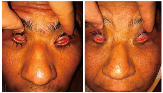
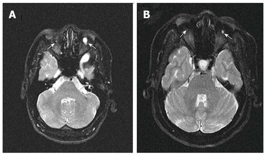
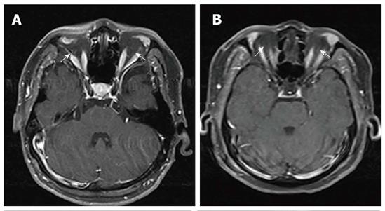
Two siblings, one a 27-year-old male (Figure 1A) and the other a 40-year-old female (Figure 1B), with anophthalmia attended the ophthalmology clinic. They were 2 of 9 siblings in a family. There was no consanguinity between their dead parents and the other siblings did not have anophthalmia or microphthalmia. Systematic physical examination and laboratory workup, including thoracoabdominal magnetic resonance imaging (MRI), were normal and the two siblings were considered to have isolated anophthalmia. Brain MRI was performed to exclude any accompanying central nervous system malformation. T1-weighted images (WI), T2-WI and post-contrast T1-WI were obtained at axial, sagittal and coronal planes. Orbital MRI was also performed to show intraorbital structures. In both cases, the globes were found to be quite small and cystic in appearance. Cornea, lens and sclera were not observed (Figure 2). Axial length of the globe was 4 mm on the right and 11 mm on the left of 27-year-old male (Figure 2A) and in the 40-year-old female, the right globe was 2 mm and the left globe was 4 mm (Figure 2B). Although both optic nerves were hypoplastic in the intraconal orbital space, the optic nerves were not found at the distal optic canal in both patients (Figure 3). In these two cases, the lacrimal glands, extraocular muscles and the eyelids were observed. Brain MRI did not show any accompanying central nervous system abnormality.
The spectrum of clinical anophthalmia includes severe microphthalmia, in which a small amount of ocular tissue can be found in the orbit on neuroimaging, even although the globe may appear to be absent externally[3]. Once the diagnosis has been established, systematic examination with both ocular and systemic imaging tests (ultrasonography, computed tomography, MRI) should be performed to rule out additional neurological, renal, cardiac or other associations[2]. Cases associated with other congenital systemic abnormalities have been reported in many syndromes, such as Fraser, Fryns, Waardenburg or Matthew-Wood[4]. Many microphthalmia patients have additional congenital ocular anomalies and associated neurological abnormalities[5]. A study conducted by Jacquemin et al[6] concluded that patients with clinical anophthalmia share a similar constellation of neurological, somatic and neuroradiological abnormalities as patients with microphthalmia. However, Schittkowski et al[7] mentioned that anophthalmia is a poorer prognostic factor than microphthalmia in terms of its association with a wide range of systemic diseases and they also described that patients with bilateral anophthalmia are characterized mainly by additional intracranial anomalies. Brunquell et al[8] described a case of anophthalmia in which severe developmental disorders and locomotor defects were recognized.
Albernaz et al[9] reported imaging findings of a clinical anophthalmia case series in which there were a variety of intraorbital, intracranial and craniofacial anomalies. The high rate of intracranial anomalies in their patients with bilateral anophthalmia suggests overall maldevelopment of the forebrain structures and failure of formation or degeneration of the optic vesicles during early gestation. MR imaging of anophthalmia cases showed amorphous tissues of isointense signal intensity with muscle on T1-WI and markedly hypointense on T2-WI. The imaging characteristics were probably due to fibrosis. These imaging features provide distinction of anophthalmia from severe microphthalmia, which is a condition of identifying a tiny globe with the normal signal intensity of the vitreous and lens[9].
However, in both of our case series, there was no syndromic condition present. Orbital and brain MRI helped us to discern the cases to be clinical anophthalmia or true anophthalmia without the need of histopathological studies. In our patients there was a bilateral hypoplastic optic nerve in the intraconal orbital space. The optic nerve was not found at the distal optic canal in both patients. There were no other central nervous system abnormalities in the brain MRI, including the posterior visual pathways. The signal intensities of rudimentary eyeballs were hypointense on T1-WI and iso-hyperintense on T2-WI due to their contents.
For depiction of the rudimentary optic nerves, optic tracts and intraorbital contents, as well as concomitant intracranial pathologies such as partial agenesis of the corpus callosum and microgyria, MRI is superior to CT[10].
In summary, we believe that the findings of orbital and brain MRI in patients diagnosed with anophthalmia has an important role in diagnosis and differentiation of cases as true anophthalmia or clinical anophthalmia.
This manuscript reported two clinical anophthalmic siblings and emphasized the importance of neuroradiological imaging findings in distinguishing true congenital anophthalmia from clinical anophthalmia.
The main limitation of the study is its nature of being a case report. More comprehensive clinical studies with anophthalmic patients are needed to emphasize the importance of neuroradiological imaging.
Anophthalmia without any systemic manifestations can be seen. Clinicians must cooperate with radiologists in cases of anophthalmia, whether true anophthalmia or severe microphthalmia.
Clinicians are strongly advised to investigate for systemic abnormalities in cases of anophthalmia. Neuroradiological imaging has an important role in diagnosing central nervous system associations with anophthalmia.
Anophthalmia is the result of insufficient development or complete regression of the optic vesicle.
The manuscript presents a rare case of two siblings with anophthalmia, demonstrating the importance of neuroradiological imaging in making the diagnosis and differentiating the case as true anophthalmia or clinical anophthalmia. It also provides a literature review regarding the intraorbital, intracranial and craniofacial anomalies associated with anophthalmia.
P- Reviewer: Battal B, Shen J, Sener RN S- Editor: Ji FF L- Editor: Roemmele A E- Editor: Lu YJ
| 1. | Verma AS, Fitzpatrick DR. Anophthalmia and microphthalmia. Orphanet J Rare Dis. 2007;2:47. [RCA] [PubMed] [DOI] [Full Text] [Full Text (PDF)] [Cited by in Crossref: 280] [Cited by in RCA: 261] [Article Influence: 14.5] [Reference Citation Analysis (0)] |
| 2. | Mafee MF. Section 2 Eye and Orbit Part 1 The Eye Pathology Congenital Anomalies. Imaging of the Head and Neck. New York: Thieme 2005; 146-147. |
| 3. | O’Keefe M, Webb M, Pashby RC, Wagman RD. Clinical anophthalmos. Br J Ophthalmol. 1987;71:635-638. [RCA] [PubMed] [DOI] [Full Text] [Cited by in Crossref: 30] [Cited by in RCA: 20] [Article Influence: 0.5] [Reference Citation Analysis (0)] |
| 4. | Makhoul IR, Soudack M, Kochavi O, Guilburd JN, Maimon S, Gershoni-Baruch R. Anophthalmia-plus syndrome: a clinical report and review of the literature. Am J Med Genet A. 2007;143:64-68. [PubMed] |
| 5. | Tucker S, Jones B, Collin R. Systemic anomalies in 77 patients with congenital anophthalmos or microphthalmos. Eye (Lond). 1996;10:310-314. [RCA] [PubMed] [DOI] [Full Text] [Cited by in Crossref: 42] [Cited by in RCA: 33] [Article Influence: 1.1] [Reference Citation Analysis (0)] |
| 6. | Jacquemin C, Mullaney PB, Bosley TM. Ophthalmological and intracranial anomalies in patients with clinical anophthalmos. Eye (Lond). 2000;14:82-87. [RCA] [PubMed] [DOI] [Full Text] [Cited by in Crossref: 10] [Cited by in RCA: 10] [Article Influence: 0.4] [Reference Citation Analysis (0)] |
| 7. | Schittkowski MP, Guthoff RF. Systemic and ophthalmological anomalies in congenital anophthalmic or microphthalmic patients. Br J Ophthalmol. 2010;94:487-493. [RCA] [PubMed] [DOI] [Full Text] [Cited by in Crossref: 21] [Cited by in RCA: 18] [Article Influence: 1.1] [Reference Citation Analysis (0)] |
| 8. | Brunquell PJ, Papale JH, Horton JC, Williams RS, Zgrabik MJ, Albert DM, Hedley-Whyte ET. Sex-linked hereditary bilateral anophthalmos. Pathologic and radiologic correlation. Arch Ophthalmol. 1984;102:108-113. [RCA] [PubMed] [DOI] [Full Text] [Cited by in Crossref: 35] [Cited by in RCA: 37] [Article Influence: 0.9] [Reference Citation Analysis (0)] |
| 9. | Albernaz VS, Castillo M, Hudgins PA, Mukherji SK. Imaging findings in patients with clinical anophthalmos. AJNR Am J Neuroradiol. 1997;18:555-561. [PubMed] |
| 10. | Daxecker F, Felber S. Magnetic resonance imaging features of congenital anophthalmia. Ophthalmologica. 1993;206:139-142. [RCA] [PubMed] [DOI] [Full Text] [Cited by in Crossref: 14] [Cited by in RCA: 14] [Article Influence: 0.4] [Reference Citation Analysis (0)] |


