Copyright
©2014 Baishideng Publishing Group Co.
World J Radiol. Apr 28, 2014; 6(4): 106-115
Published online Apr 28, 2014. doi: 10.4329/wjr.v6.i4.106
Published online Apr 28, 2014. doi: 10.4329/wjr.v6.i4.106
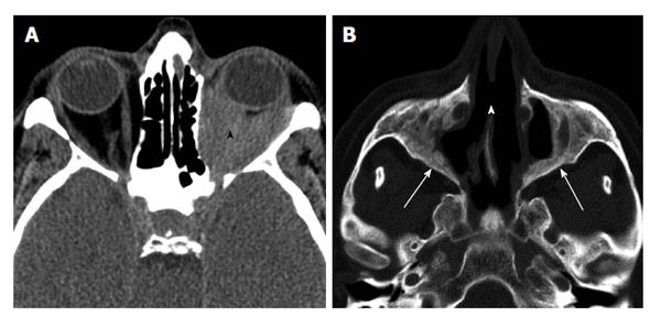
Figure 11 Wegener's granulomatosis.
A: Non-enhanced axial CT through orbit demonstrating diffuse infiltration of orbital fat (black arrowhead); B: Axial CT in through sinuses in bone window shows destruction of medial maxillary sinus walls, perforation of nasal septum (white arrowhead), and chronic neo osteogenesis along sinus walls (white arrows). CT: Computed tomography.
- Citation: Pakdaman MN, Sepahdari AR, Elkhamary SM. Orbital inflammatory disease: Pictorial review and differential diagnosis. World J Radiol 2014; 6(4): 106-115
- URL: https://www.wjgnet.com/1949-8470/full/v6/i4/106.htm
- DOI: https://dx.doi.org/10.4329/wjr.v6.i4.106


