Published online Nov 28, 2014. doi: 10.4329/wjr.v6.i11.886
Revised: September 20, 2014
Accepted: October 1, 2014
Published online: November 28, 2014
Processing time: 162 Days and 11.8 Hours
Truncus arteriosus is an uncommon congenital cardiac abnormality which is characterized by a single arterial trunk origin from the heart that supplies both the systemic, pulmonary and coronary circulation. We present a preterm newborn female patient with type 2 truncusarteriosus, left superior vena cava and aberrant subclavian artery diagnosed with low dose dual-source cardiac computed tomography (CT). We discuss that low dose dual-source cardiac CT has more advantages than other imaging methods and it is an important modality for assessment of patients with conotruncal anomalies such as truncusarteriosus.
Core tip: Truncus arteriosus is an uncommon congenital cardiac abnormality which is characterized by a single arterial trunk origin from the heart that supplies both the systemic, pulmonary and coronary circulation. We discuss that low dose dual-source cardiac computed tomography has more advantages than other imaging methods and it is an important modality for assessment of patients with conotruncal anomalies such as truncusarteriosus.
- Citation: Koplay M, Cimen D, Sivri M, Güvenc O, Arslan D, Nayman A, Oran B. Truncus arteriosus: Diagnosis with dual-source computed tomography angiography and low radiation dose. World J Radiol 2014; 6(11): 886-889
- URL: https://www.wjgnet.com/1949-8470/full/v6/i11/886.htm
- DOI: https://dx.doi.org/10.4329/wjr.v6.i11.886
Truncus arteriosus is an uncommon congenital cardiac abnormality which is characterized by a single arterial trunk origin from the ventricle which occurs due to the failure of conotruncalseptation during development of the fetus. It occurs in approximately 2% of all congenital cardiac anomalies and it is seen higher in males than females[1]. This arterial trunk enables systemic, pulmonary, and coronary circulation. In general, the common trunk is combined with a large, sub arterial ventricular septal defect (VSD) of infundibular type to providethe completion of circulatory flow circuit[2]. Less often, it may originate completely right or left ventricle[3]. Several different abnormalities are described with truncusarteriosus that lead to differences in diagnosis and treatment such as the interruption of aortic arch, structural abnormalities of the truncal valve, coronary artery abnormalities, and much more rarely, right aortic arch, double aortic arch, left superior vena cava, secundum atrial septal defect (ASD), aberrant subclavian artery and complete atrioventricular septal defect. Prenatal ultrasound, echocardiography, catheter cardiac angiography, computerized tomography (CT) and magnetic resonance imaging (MRI) can be used for diagnosis of congenital cardiovascular diseases[4]. The purpose of this study was to present a case of TA arising from completely right ventricle with left superior vena cava and aberrant subclavian artery and to describe the advantages of low dose dual-source CT angiography in diagnosis.
A three-day preterm female infant was referred to our clinic with cleft lip-palate and on suspicion of cardiac abnormality. She was born at 36th week of gestation by caesarian delivery with the birth-weight of 2600 g from a 33-year-old woman. Physical examination showed central cyanosis and grade 2/6 systolic murmur at the apex. Respiratory rate was 48 breaths/min, and heart rate was 160 beats/min. The lungs found clear by auscultation and the liver was palpable 4 cm under the right costal margin. Arterial blood gas results revealed that PaO2 was 40.6 mmHg, PaCO2 as 16.9 mmHg, and O2SAT as 84%. Transthoracic echocardiography showed the secundum type ASD, peri-membraneous VSD, and single arterial trunk.
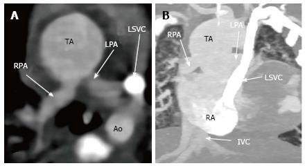
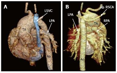
In order to demonstrate such congenital anomalies and vascular structures in detail, multi-detector computerized tomography (MDCT) was used on a dual-source 128-MDCT scanner (Somatom Definition Flash, Siemens Healthcare, Germany). No medication was administered for sedation. Scans were acquired by 128 mm × 0.625 mm collimation, 3 mm slice thickness, 0.6 mm reconstruction slice thickness, and 0.3 mm reconstruction interval, 80 kVp, 25 mA and a helical pitch of 3.4. Non-ionic contrast medium (1.5 mL/kg) was applied by an automatic injector at a rate of 1 mL/s. CT scan was obtained from the arcus aorta level towards the diaphragmatic face of the heart in prospectively electrocardiography (ECG)-triggered high-pitch spiral mode (flash spiral technique). After the traditional images of the patient were acquired on axial plane and were evaluated in detail. In addition, multiplanar reconstructions (MPR) maximum intensity projection (MIP) and Three dimensional (3D) volume rendering (3D VR) images were used for evaluating of the anomalies by using special computerized software (Syngovia, 2011). MDCT showed secundum type ASD and peri-membraneous VSD as echocardiography. Additionally, there was a single arterial trunk origin from the right ventricle. Left and right branched pulmonary arteriesweredivided into the posterior aspect of trunk. Furthermore, persistent left superior vena cava and right aberrant subclavian artery were diagnosed during the CT study. 3D VR images clearly demonstrated the correlation between these abnormal vessels and origins (Figures 1 and 2). The patient died due to bronchopneumonia.
The radiation dose was determined in terms of protocol dose-length product (DLP) in CT scanning. Effective dose (ED) was obtained on the value of DLP multiplying it by 0.039 conversion factor for infant. The DLP value for CT angiography was 10 mGy cm and estimated ED was calculated as 0.39 mSv.
Truncus arteriosus is a major conotruncal anomaly such as the Fallot tetralogy, double-outlet right ventricle, transposition of the great vessels and interrupted arcus aorta. It is related with chromosome 22q11 deletion and DiGeorge syndrome. Two classifications have been identified: one by Collett and Edwards in 1949 and the other one by Van Praagh[4] in 1965. There are four types of truncus arteriosus based on the branching pattern of pulmonary artery in each classification system. In Collets and Edwards classification, there is a single pulmonary trunk which origins from the left lateral aspect of the common trunk and pulmonary trunk was divided into right and left pulmonary arteries in type 1. In type 2, pulmonary trunk is absent and right and left pulmonary arterial branches origin from the posterolateral aspect of the common arterial trunk as in our case. Type 3, left and right pulmonary arteries origin from the left and right lateral aspects of the trunk. Type 4, major aorta-pulmonary collateral arteries enables pulmonary blood flow. Van Praagh classification is nearly the same the classification of Collett and Edwards. There are some similar differences.
In diagnosis, chest radiography may be the first simplest technique to show cardiomegaly and increased pulmonary vascular markings. However, it usually does not provide detailed diagnosis.
Echocardiography is a basic, rapid, non-radiating and non-invasive method for the diagnosis of TA. It may lead to determine hemodynamics. However, there are some limitations of echocardiography such as a small field of view (FOV) and an acoustic window. Also it is operator-dependent and the image quality is less in geriatric patients. It is also inadequate for visualization of anomalous vessel anatomy, origin and branching of arterial trunk, other associated anomalies with TA, and extra cardiacstructures[5].
Cardiac catheterization and angiography can be used for interventional procedures. It is now used less frequently for the diagnosis because it is an invasive method and it requiressedation or general anesthesia. Furthermore, it has catheter-related complications and causes the patient expose to high radiation doses and iodine-containing contrast agent.
Cardiovascular MRI is one of the best modalities for the diagnosis of TA. Being non-invasive and non-radiating, cardiovascular MRI provides structural and functional information such as ventricular volumes and function, flow in chambers and vessels, and tissue characteristics[5]. Disadvantages of MRI are that, it is less accessible and is expensive. It can be difficult for the patients to stay still for a long scanning time. Sedation or general anesthesia is required in young patients. Vascular stents, coils, and pacemakers can cause metallic artefacts.
CT provides excellent morphological evaluation of TA. 3D CT angiography provides greater information about anomalous anatomic detail, abnormal origin, branching of arterial trunk, cardiac and extra cardiac abnormalities such asaortic arch, coronary arteries abnormalities, left superior vena cava, and aberrant subclavian artery[5,6]. 3D images provide our understanding of complex anatomy and connection problems. Additionally, it is useful for surgical planning and post-operative assessment. It is easy to use especially in younger patients due to the fast acquisition time and necessity of minimal sedation[5,7]. It is more practical compared to MRI. Furthermore, CT can be safely used in patients with vascular stents, coils, and pacemakers. Airways and lung parenchyma can be evaluated simultaneously[7].
The significant disadvantages of CT when compared to MRI are ionizing radiation and iodinated contrast media, to which infants and children are especially sensitive. Moreover pediatric patients normally have higher heart rates and may have an incompatibility against beta-blockers. Breath-holding is an important problem in pediatric patients, as well. Flash spiral mode of dual-source CT can be used to overcome these disadvantages. Dual Source CT (DSCT) technology is the latest innovation in MDCT. In DSCT, ionizing radiation and the necessity of contrast media can be minimized with the usage of a weight-based low-dose protocol. Moreover, it allows high temporal resolution in patients with high heart rates or arrhythmia and does not require the use of beta-blockers. Providing high image quality in a shortest breah-holding period, as well, this technology is a fast scanning method due to the application of dual X-ray and detector system simultaneously[8]. They are very important features for pediatric cardiac CT examinations of congenital heart diseases. In our case, DSCT have clearly showed findings indicative of type 2 TA, the left and right branched pulmonary arteries that arose from posterior aspect of trunk. The ionizing radiation dose and contrast volume were calculated as 0.39 mSv and 4 mL, respectively.
In conclusion, DSCT is a useful imaging method for diagnosis, surgical planning, and postoperative evaluation of congenital heart abnormality such as TA, especially in infants and in children. It has significant roles to get the better limitations of other imaging modalities and should be preferred because of its fast imaging quality, low radiation dose, short breath-hold, and the other advantages.
A three day preterm female infant was referred to our clinic with cleft lip -palate and suspicion of cardiac anomaly.
Physical examination showed central cyanosis and grade 2/6 systolic murmur at the apex.
Cardiovascular anomalies.
Arterial blood gas results revealed that PaO2 was 40.6 mmHg, PaCO2 of 16.9 mmHg, O2SAT of 84%.
Multidedector computer tomography (MDCT) showed secundum type atrial septal defect and perimembraneous ventricular septal defect as echocardiography; in additionally, there was a single arterial trunk arising from the right ventricule, persistent left superior vena cava and right aberrant subclavian artery was diagnosed on CT study.
Truncus arteriosus.
The patient was died due to bronchopneumonia.
CT is a useful imaging method for diagnosis, surgical planning and postoperative evaluation of congenital heart diseases like truncus arteriosus especially in infants and children.
Dual source CT systems have design of a CT scanner with two X-ray tubes and two detectors that has the potential to overcome limitations of conventional MDCT systems, such as temporal resolution for cardiac imaging.
Dual-source CT has significant roles to get the better limitations of other imaging modalities and should be preferred because of its fast imaging quality, low radiation dose, short breath-hold and the other advantages.
The manuscript is well written.
P- Reviewer: Battal B, Cademartiri F, Hiwatashi A S- Editor: Ji FF L- Editor: A E- Editor: Lu YJ
| 1. | Pilu G, Nicolaides KH. Diagnosis of Fetal Abnormalities: The 18-23-Week Scan. New York, NY: Taylor and Francis 1999; . |
| 2. | Barboza JM, Dajani NK, Glenn LG, Angtuaco TL. Prenatal diagnosis of congenital cardiac anomalies: a practical approach using two basic views. Radiographics. 2002;22:1125-1137; discussion 1125-1137. [RCA] [PubMed] [DOI] [Full Text] [Cited by in Crossref: 37] [Cited by in RCA: 38] [Article Influence: 1.7] [Reference Citation Analysis (0)] |
| 3. | Murdison KA, McLean DA, Carpenter B, Duncan WJ. Truncusarteriosuscommunis associated with mitral valve and left ventricular hypoplasia without ventricular septal defect: unique combination. Pediatr Cardiol. 1996;17:322-326. [RCA] [PubMed] [DOI] [Full Text] [Cited by in Crossref: 18] [Cited by in RCA: 18] [Article Influence: 0.6] [Reference Citation Analysis (0)] |
| 4. | Yildirim A, Karabulut N, Doğan S, Herek D. Congenital thoracic arterial anomalies in adults: a CT overview. DiagnInterv Radiol. 2011;17:352-362. [PubMed] |
| 5. | Johnson TR. Conotruncal cardiac defects: a clinical imaging perspective. Pediatr Cardiol. 2010;31:430-437. [RCA] [PubMed] [DOI] [Full Text] [Cited by in Crossref: 21] [Cited by in RCA: 22] [Article Influence: 1.5] [Reference Citation Analysis (0)] |
| 6. | Kawano T, Ishii M, Takagi J, Maeno Y, Eto G, Sugahara Y, Toshima T, Yasunaga H, Kawara T, Todo K. Three-dimensional helical computed tomographic angiography in neonates and infants with complex congenital heart disease. Am Heart J. 2000;139:6546-6560. [RCA] [DOI] [Full Text] [Cited by in Crossref: 37] [Cited by in RCA: 30] [Article Influence: 1.2] [Reference Citation Analysis (0)] |
| 7. | Goo HW, Park IS, Ko JK, Kim YH, Seo DM, Yun TJ, Park JJ, Yoon CH. CT of congenital heart disease: normal anatomy and typical pathologic conditions. Radiographics. 2003;23 Spec No:S147-S165. [RCA] [PubMed] [DOI] [Full Text] [Cited by in Crossref: 175] [Cited by in RCA: 137] [Article Influence: 6.2] [Reference Citation Analysis (0)] |
| 8. | Matt D, Scheffel H, Leschka S, Flohr TG, Marincek B, Kaufmann PA, Alkadhi H. Dual-source CT coronary angiography: image quality, mean heart rate, and heart rate variability. AJR Am J Roentgenol. 2007;189:567-573. [RCA] [PubMed] [DOI] [Full Text] [Cited by in Crossref: 148] [Cited by in RCA: 127] [Article Influence: 7.1] [Reference Citation Analysis (0)] |


