Copyright
©2012 Baishideng Publishing Group Co.
World J Radiol. Aug 28, 2012; 4(8): 345-352
Published online Aug 28, 2012. doi: 10.4329/wjr.v4.i8.345
Published online Aug 28, 2012. doi: 10.4329/wjr.v4.i8.345
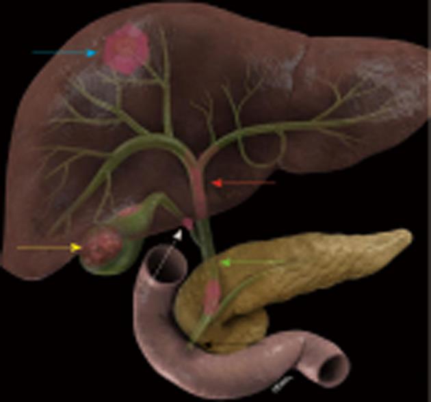
Figure 1 Anatomical distribution of cholangiocarcinoma.
The intrahepatic cholangiocarcinoma present most as solid masses in the liver (blue arrow), gallbladder (GB) cancer present as solid mass in the GB (yellow arrow), proximal bile duct tumors present mostly as infiltrating masses (red arrow), middle bile duct tumors (green arrow), and distal bile duct tumors (black arrow). The cystic duct is labeled with a white arrow.
- Citation: Ganeshan D, Moron FE, Szklaruk J. Extrahepatic biliary cancer: New staging classification. World J Radiol 2012; 4(8): 345-352
- URL: https://www.wjgnet.com/1949-8470/full/v4/i8/345.htm
- DOI: https://dx.doi.org/10.4329/wjr.v4.i8.345


