Published online May 28, 2012. doi: 10.4329/wjr.v4.i5.231
Revised: December 12, 2011
Accepted: December 19, 2011
Published online: May 28, 2012
Right-sided aortic arch with aberrant left subclavian artery (RAA/ALSC) is the second most common mediastinal complete vascular ring. Adult presentation of dysphagia lusoria due to a RAA/ALSC is uncommon with fewer than 25 cases reported in the world literature. The left lateral portion of this vascular ring is not a vessel, but an atretic ductus arteriosus, the ligamentum arteriosum, which has been identified in different cases as the major cause of tracheo-esophageal impingement. Surgical division of the ligamentum arteriosum allows the vessels to assume a less constricting pattern decreasing dysphagic symptoms. Clear visualization of the ligamentum arteriosum by diagnostic imaging has not been obtained in previously reported cases. We demonstrated, using magnetic resonance imaging, the location and the complete course of a left-sided ligamentum arteriosum in a patient with adult-onset dysphagia due to a RAA/ALSC with a small Kommerell’s diverticulum, providing, during the same session, a complete assessment of both mediastinal vascular abnormalities and esophageal impingement sites.
- Citation: Paparo F, Bacigalupo L, Melani E, Rollandi GA, De Caro G. Cardiac-MRI demonstration of the ligamentum arteriosum in a case of right aortic arch with aberrant left subclavian artery. World J Radiol 2012; 4(5): 231-235
- URL: https://www.wjgnet.com/1949-8470/full/v4/i5/231.htm
- DOI: https://dx.doi.org/10.4329/wjr.v4.i5.231
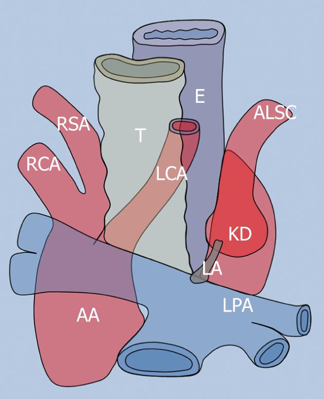
Right-sided aortic arch with aberrant left subclavian artery (RAA/ALSC) is the second most common mediastinal complete vascular ring occurring in an estimated 1 in 1000 individuals in the general population. This type of vascular ring has a 10% association with intracardiac defects[1] (Figure 1).
A RAA may be asymptomatic. Adult-onset dysphagic symptoms are thought to be the result of early atherosclerotic changes of the anomalous vessels, dissection, or aneurysmal dilatation with compression of surrounding mediastinal structures, including the esophagus[1-6]. Adult presentation of dysphagia lusoria due to a RAA/ALSC is uncommon with fewer than 25 cases reported in the world literature[2]. The left lateral portion of this vascular ring is not a vessel, but an atretic ductus arteriosus, the ligamentum arteriosum, which has been identified in different cases as the major cause of tracheo-esophageal impingement[1-4]. The most common approach to a RAA/ALSC vascular ring is a left posterolateral thoracotomy which provides the best access to the middle and posterior mediastinum. Surgical division of the ligamentum arteriosum with dissection of mediastinal structures allows the vessels to assume a less constricting pattern and division of the retroesophageal artery is often unnecessary[1-4]. Cross-sectional diagnostic imaging techniques [magnetic resonance imaging (MRI), MR-angiography and MDCT-angiography] and digital angiography may provide complete visualization of vascular rings, but a clear demonstration of the ligamentum arteriosum has not been obtained in previously reported cases[2-5].
We demonstrated, using MRI, the location and the complete course of a left-sided ligamentum arteriosum in a patient with adult-onset dysphagia due to a RAA/ALSC with a small Kommerell’s diverticulum, providing, during the same session, a complete demonstration of both mediastinal vascular abnormalities and esophageal impingement sites.
A 74-year-old man presented with 1-year history of progressive dysphagia, first to solids, then to semisolids. His physical exam was unremarkable and laboratory values were normal. His medical history was negative for respiratory and cardiac diseases. Chest radiography revealed an abnormality of the aortic arch and descending thoracic aorta, which was interpreted as a RAA with a right descending thoracic aorta, and the diagnosis of a tracheo-esophageal compression syndrome due to a vascular ring was entertained. The patient underwent an MRI examination (Signa™ HDxt 1.5T, GE Healthcare) including three consecutive parts:
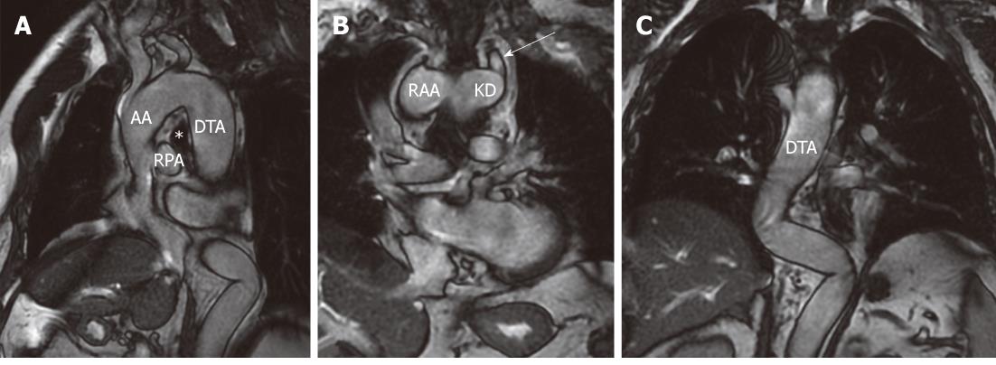
The first vessel originating from the RAA (at the passage between ascending aorta and RAA) was the left carotid artery, which travelled anterior to the trachea. The RAA then gave off the right carotid followed by the right subclavian artery. The ALSC originated from a Kommerell’s diverticulum, which represents the non-resorbed remnant of the embryonic left fourth arch, placed at the point of merger between the RAA and proximal descending thoracic aorta. The distal descending thoracic aorta was seen on the right-side of the spine and it had an abrupt angulation to the left, just proximal to the diaphragmatic hiatus (Figure 2).
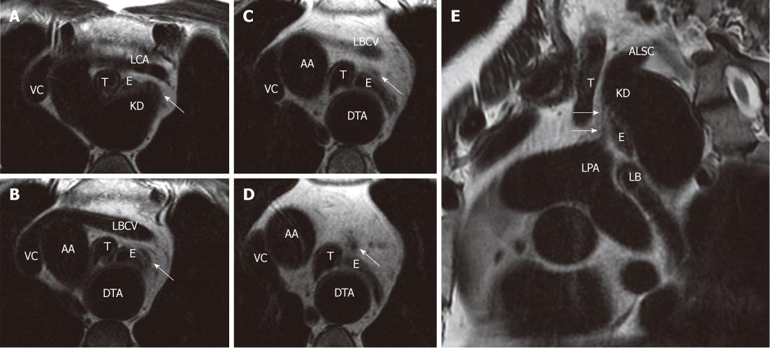
Demonstration of location and course of ligamentum arteriosum and its relationships with the tracheo-esophageal bundle (cardiac-gated frFSE T2 weighted sequence on axial and sagittal oblique planes)
The ligamentum arteriosum (non-functional vestige of the ductus arteriosus) arose from the base of Kommerell’s diverticulum, just inferiorly from the origin of the ALSC, and attached distally to the superior aspect of the left pulmonary artery. The trachea and esophagus were surrounded by the left common carotid artery anteriorly, ascending aorta and proximal part of the aortic arch on the right side, retroesophageal part of the RAA posteriorly, Kommerell’s diverticulum and left ligamentum arteriosum on the left posterolateral and left lateral sides, respectively. The entire course of the ligamentum was demonstrated by a sagittal oblique frFSE T2-weighted sequence (Figure 3).
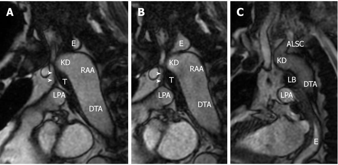
The field of view of the cine-MRI sequence was placed to simultaneously visualize the cervical and thoracic esophageal segments, and distension of the esophageal lumen was evaluated (Figure 4). This sequence has a short acquisition time and it may detect fluids in motion, such as a bolus of water, providing a series of image frames.
During dynamic evaluation of the esophageal swallowing phase a progressive distension of the esophageal tract superior to the RAA was noticed.
The water bolus was subsequently appreciated in the esophageal lumen inferiorly to the proximal left main bronchus. The dynamic sagittal oblique FIESTA sequence was then repeated in an attempt to obtain distention of the mid-portion of the thoracic esophagus passing through the vascular ring; however, it only demonstrated a narrow passage for water without a significant distension of the esophageal lumen.
Dysphagia lusoria is used to describe symptomatic extrinsic compression of the esophagus from any vascular abnormality of the aortic arch, and was first described by Bayford in 1787, who introduced the term “arteria lusoria” (after lusus naturae).
The most common embryologic abnormality of the aortic arch related to esophageal compression and dysphagia lusoria is an aberrant right subclavian artery, which occurs in 0.5%-1.8% of the population. RAA, with or without an ALSC, is the second most common cause of dysphagia lusoria[6-8].
Our patient had a type II RAA accordingly to Edwards[7] (RAA with ALSC) with a left ligamentum arteriosum. This rare complete vascular ring has been well described in the medical literature and represents 39.5% of all right aortic arches. It is largely asymptomatic with 75% of symptomatic patients occurring mainly in infancy and early childhood. Adult-onset dysphagia lusoria is thought to be the consequence of fibrous transformation of paratracheal and paraesophageal connective tissues together with an age-related atheromatous process[3-5]. The presence of an aneurysm of the ALSC or Kommerell’s diverticulum may represent another cause of dysphagic symptoms in these patients[7].
In type II RAA, Kommerell’s diverticulum represents the broad base from which ALSC takes off and it also offers proximal attachment to the left ligamentum arteriosum, which is considered the left lateral aspect of the vascular ring[1-4].
In adults, division of the ligamentum with dissection of mediastinal structures allows the vessels to assume a less constricting pattern, and surgical division of ALSC is frequently avoided[1-5]. The exact location and course of ligamentum arteriosum have never been depicted by MRI in previously reported cases. We used MRI to demonstrate superior mediastinal vascular anatomy and the relationships between abnormal mediastinal vessels, ligamentum arteriosum and tracheo-esophageal bundle in our patient.
Dynamic evaluation of the esophageal swallowing phase demonstrated two main impingement sites along the esophageal course, the upper site was related to the retroesophageal part of the RAA, the inferior site was due to the left lateral compression by the ligamentum arteriosum.
Cine-MRI did not reveal severe stops along the esophageal course, but it was impossible to appreciate distension of the mid thoracic esophagus passing through the vascular ring.
These imaging findings showed good correlation with the mild-moderate dysphagic symptoms in our patient (grade 2 dysphagia[9]).
Cine-MRI has not been previously used to evaluate esophageal motility, and its ability to demonstrate esophageal impingement sites requires to be assessed by more extensive studies. In this case cine-MRI was very useful, providing good visualization of the anatomical structures surrounding the esophagus, which are not appreciable with conventional barium studies.
The correct diet allowed the symptoms to be controlled and the patient had an uneventful 12-mo follow-up period. The patient’s conservative treatment did not allow surgical correlation of MRI findings, and this probably represents the most important drawback of our report.
However, the hypointense band-shaped element we identified on a cardiac-gated MRI sagittal oblique sequence (Figure 3) reflects the exact location and course of the ligamentum arteriosum, as described in cadaveric specimens, surgical procedure photographs and anatomical tables and drawings[1-5].
The work-up of an adult patient with dysphagia, besides a high index of suspicion, includes standard chest radiography, barium esophagogram, esophageal manometry and esophageal fiber-optic endoscopy. Cross-sectional imaging techniques (MDCT, digital subtraction angiography, MRI and MRA)[3-5,9] are used to depict vascular rings and aberrant vessels in dysphagia lusoria.
Plain film chest radiography and barium esophagogram are often the first approach in dysphagic patients. However, these imaging modalities provide only indirect findings and limited data. Barium esophagogram and esophageal fiber-optic endoscopy may demonstrate luminal narrowing due to extrinsic esophageal compression with an intact mucosa, excluding other causes of dysphagia[3-6]. Manometric investigation reveals nonspecific abnormalities[9]. Digital subtraction angiography provides complete and detailed information regarding mediastinal vascular anatomy, however, extravascular structures are not visualized. MDCT angiography is an established diagnostic imaging technique in the evaluation of many chest vascular diseases and it may demonstrate the relationships between abnormal vessels and the tracheo-esophageal bundle[6], but the ligamentum arteriosum is not appreciable on CT images[4,6].
The correct detection of anatomical structures responsible for esophageal impingement sites has a relevant clinical utility, and good visualization of the complete course of the ligamentum arteriosum may be useful for the surgical planning of its division in patients with more severe dysphagic symptoms.
We believe that MRI may represent an all-in-one diagnostic tool to investigate dysphagia lusoria avoiding both radiation exposure and the use of paramagnetic and iodinated intravenous contrast agents.
Peer reviewers: Edwin JR van Beek, MD, PhD, Med, FRCR, SINAPSE, Chair of Medical Imaging, Clinical Imaging Research Centre, CO.19, Queen’s Medical Research Institute, 47 Little France Crescent, Edinburgh EH16 4TJ, United Kingdom; Dr. Markus Weininger, Department of Radiology, University Hospital of Wuerzurg, Auf der Laeng 24, 97076 Wuerzburg, Germany
S- Editor Cheng JX L- Editor Webster JR E- Editor Zheng XM
| 1. | Morris CD, Kanter KR, Miller JI. Late-onset dysphagia lusoria. Ann Thorac Surg. 2001;71:710-712. [RCA] [PubMed] [DOI] [Full Text] [Cited by in Crossref: 10] [Cited by in RCA: 13] [Article Influence: 0.5] [Reference Citation Analysis (0)] |
| 2. | Sitzman TJ, Mell MW, Acher CW. Adult-onset dysphagia lusoria from an uncommon vascular ring: a case report and review of the literature. Vasc Endovascular Surg. 2009;43:100-102. [RCA] [PubMed] [DOI] [Full Text] [Cited by in RCA: 1] [Reference Citation Analysis (0)] |
| 3. | Koullias GJ, Korkolis DP, Iams WB, Elefteriades JA. Late-onset dysphagia lusoria assessed by 3-dimensional computed tomography of an aortic arch abnormality. Dis Esophagus. 2005;18:60-63. [RCA] [PubMed] [DOI] [Full Text] [Cited by in Crossref: 1] [Cited by in RCA: 2] [Article Influence: 0.1] [Reference Citation Analysis (0)] |
| 4. | Zhao J, Liao Y, Gao S. Right aortic arch with retroesophageal left ligamentum arteriosum. Tex Heart Inst J. 2006;33:218-221. [PubMed] |
| 5. | Bashar AH, Kazui T, Yamashita K, Terada H, Washiyama N, Suzuki K. Right aortic arch with aberrant left subclavian artery symptomatic in adulthood. Ann Vasc Surg. 2006;20:529-532. [RCA] [PubMed] [DOI] [Full Text] [Cited by in Crossref: 2] [Cited by in RCA: 1] [Article Influence: 0.1] [Reference Citation Analysis (0)] |
| 6. | Alper F, Akgun M, Kantarci M, Eroglu A, Ceyhan E, Onbas O, Duran C, Okur A. Demonstration of vascular abnormalities compressing esophagus by MDCT: special focus on dysphagia lusoria. Eur J Radiol. 2006;59:82-87. [RCA] [PubMed] [DOI] [Full Text] [Cited by in Crossref: 26] [Cited by in RCA: 28] [Article Influence: 1.5] [Reference Citation Analysis (0)] |
| 7. | Cinà CS, Althani H, Pasenau J, Abouzahr L. Kommerell's diverticulum and right-sided aortic arch: a cohort study and review of the literature. J Vasc Surg. 2004;39:131-139. [RCA] [PubMed] [DOI] [Full Text] [Cited by in Crossref: 254] [Cited by in RCA: 267] [Article Influence: 12.7] [Reference Citation Analysis (0)] |
| 8. | Adkins RB, Maples MD, Graham BS, Witt TT, Davies J. Dysphagia associated with an aortic arch anomaly in adults. Am Surg. 1986;52:238-245. [PubMed] |
| 9. | Janssen M, Baggen MG, Veen HF, Smout AJ, Bekkers JA, Jonkman JG, Ouwendijk RJ. Dysphagia lusoria: clinical aspects, manometric findings, diagnosis, and therapy. Am J Gastroenterol. 2000;95:1411-1416. [RCA] [PubMed] [DOI] [Full Text] [Cited by in Crossref: 97] [Cited by in RCA: 88] [Article Influence: 3.5] [Reference Citation Analysis (0)] |


