Published online Nov 28, 2011. doi: 10.4329/wjr.v3.i11.273
Revised: August 7, 2011
Accepted: August 14, 2011
Published online: November 28, 2011
AIM: To investigate the features of crush thoracic trauma in Sichuan earthquake victims using chest digital radiography (CDR).
METHODS: We retrospectively reviewed 772 CDR of 417 females and 355 males who had suffered crush thoracic trauma in the Sichuan earthquake. Patient age ranged from 0.5 to 103 years. CDR was performed between May 12, 2008 and June 7, 2008. We looked for injury to the thoracic cage, pulmonary parenchyma and the pleura.
RESULTS: Antero-posterior (AP) and lateral CDR were obtained in 349 patients, the remaining 423 patients underwent only AP CDR. Thoracic cage fractures, pulmonary contusion and pleural injuries were noted in 331 (42.9%; 95% CI: 39.4%-46.4%), 67 and 135 patients, respectively. Of the 256 patients with rib fractures, the mean number of fractured ribs per patient was 3. Rib fractures were mostly distributed from the 3rd through to the 8th ribs and the vast majority involved posterior and lateral locations along the rib. Rib fractures had a significant positive association with non-rib thoracic fractures, pulmonary contusion and pleural injuries (P < 0.001). The number of rib fractures and pulmonary contusions were significant factors associated with patient death.
CONCLUSION: Earthquake-related crush thoracic trauma has the potential for multiple fractures. The high number of fractured ribs and pulmonary contusions were significant factors which needed appropriate medical treatment.
- Citation: Dong ZH, Shao H, Chen TW, Chu ZG, Deng W, Tang SS, Chen J, Yang ZG. Digital radiography of crush thoracic trauma in the Sichuan earthquake. World J Radiol 2011; 3(11): 273-278
- URL: https://www.wjgnet.com/1949-8470/full/v3/i11/273.htm
- DOI: https://dx.doi.org/10.4329/wjr.v3.i11.273
An earthquake with a magnitude of 8.0 which occurred on May 12, 2008 struck Sichuan province in China and the earthquake activity was felt by the Chinese people from Gansu to Liaoning provinces. According to official reports, approximately 374 643 people were injured, 69 227 people died and 17 923 were missing at 12:00 on September 25, 2008[1]. Our major university hospital located 92 kilometers away from the epicenter, Wenchuan County, was an important rescue center which was undamaged. This hospital, the nearest and biggest in the southwest region of China, treated a total of 2674 wounded patients (869 out-patients; 1805 in-patients) by July 7, 2008. Patients were conveyed to the hospital mainly by vehicles or helicopter.
Chest injuries comprised 7.6%-15.9% of all earthquake-related hospitalized patients[2-4]. In the Marmara earthquake, the mortality of patients with a major chest injury was up to 16% (three of 19 patients). In patients who had suffered coexistent trauma with chest injury and with an injury severity score over 25, mortality may reach 60%[5]. As the injured must be retrieved from beneath collapsed rubble, earthquake-related thoracic injuries still present a major challenge to emergency medical staff[6]. In our previous study, we found that crush thoracic trauma arising from the Sichuan earthquake was life threatening, and had different multidetector computed tomography characteristics compared with other reported common thoracic traumas[7]. Therefore, it was crucial to timely detect this type of chest injury to ensure appropriate emergency medical treatment. In order to identify possible chest injuries quickly, an antero-posterior (AP) chest radiograph remains the most common first imaging method obtained in these patients[8,9]. A large data sample of the chest digital radiography (CDR) features of crush chest trauma in earthquake victims has not been reported[2-4]. Thus, we retrospectively reviewed the CDR appearance of 772 consecutive patients with clinical crush thoracic trauma resulting from the Sichuan earthquake to investigate the features of crush thoracic trauma in Sichuan earthquake victims using CDR, and to evaluate the use of CDR in such a disaster.
Of the 2674 patients treated in our hospital, a significant number had sustained crush injuries due to building collapse, flying stones, bricks or other falling objects. Chest CDR were ordered by clinicians and performed on patients with thoracic trauma. Patients were enrolled into our study according to the following criteria: (1) the etiology of the injury was associated with the 2008 Sichuan earthquake; (2) thoracic injuries were evaluated by CDR; and (3) the patients had not received related surgical therapies before CDR. Patients were excluded from the present study according to the following criteria: (1) patients who jumped or accidentally fell from heights due to the earthquake; (2) patients who were injured by earthquake-related motor vehicle injury, burn injuries or other etiologies; and (3) patients who received CDR routinely preoperatively due to non-thoracic injuries.
CDR was performed using a digital radiographic system (Digital Diagnost, Philips Healthcare, Hamburg, Germany) with a flat-panel detector. Because some patients had severe injuries to thoracic or non-thoracic organs, only 349 patients could complete AP and lateral CDR, the remaining 423 patients underwent only AP CDR, 387 of which were supine AP CDR. For the AP images, the following parameters were used: 90 kV, 1-12 mAs, an active imaging area of 43 cm × 43 cm, theoretical spatial resolution of 3.5 line pairs per millimeter, and a matrix of 3001 × 3001 pixels. As for the lateral images, the parameters were similar to those used for AP images except for a tube tension of 109 kV and tube current of 3-28 mAs.
The following information was determined by consensus between two radiologists: (1) number of fractured ribs; (2) the location of rib fracture(s) along the rib (anterior, lateral, or posterior); and (3) non-rib thoracic fractures including sternum, clavicle, scapula and thoracic spinal fractures. Patients were also analyzed according to pulmonary contusion, pleural injuries, and adjacent cervical and lumbar spinal fractures (C6, C7, L1 and L2 vertebra fractures). As the distinction between pulmonary condensations and contusion is difficult, for the diagnosis of pulmonary contusion, we excluded patients who had a fever, or who had acute renal failure due to crush syndrome, or who received CDR 14 d after the crush injuries.
Data analysis was performed on a personal computer with the SPSS statistical package (version 13.0 for Windows, SPSS Inc., Chicago, IL, USA). The Chi-square test was used to evaluate the relationships between rib fractures and non-rib thoracic fractures, pulmonary contusion and pleural injuries. The Wilcoxon W test was used to compare the number of fractured ribs between patients with and without non-rib thoracic fractures. Multivariate logistic regression analysis was performed to assess the risk factors in this cohort. The patients were sorted into two clinical outcome groups [patients who had a fatal outcome (the “fatal” group) and patients who were discharged alive from our hospital (the “discharged” group)]. We collected data on two dichotomous variables: pulmonary contusion and pleural injury, and two ordered categorical variables: age (< 35 years, 35-64 years, and ≥ 65 years of age) and number of fractured ribs (0, 1-2, and ≥ 3). Logistic regression was performed to assess these risk factors. A two-tailed P-value of less than 0.05 wasaccepted as a statistically significant
difference.
Between May 12, 2008 and June 7, 2008, a total of 772 patients (28.87% of the 2674 patients who presented to our hospital) suffered crush thoracic trauma in the earthquake and were reviewed. The main clinical findings in these patients were chest pain, cough, hemoptysis, and dyspnea. The mean time from injury to CDR was 6 d, with a range of 30 min to 25 d. There were 355 male patients (age range, 2.5 to 90 years; median age, 49 years) and 417 female patients (age range, 0.5 to 103 years; median age, 55 years). The median age of the total cohort was 51 years.
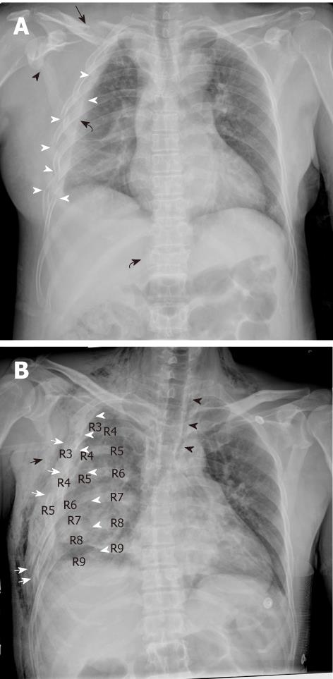
Of the 772 patients, thoracic injury occurred in 353 cases (45.7%; 13.2% of these 2674 patients). Fractures were detected in 331 patients (42.9%) including rib fractures in 256 patients (33.2%), non-rib thoracic fractures in 115 patients, and adjacent cervical and lumbar spinal fractures in 54 patients. Pulmonary contusion was determined in 67 patients (8.7%). Pleural injuries including hemothorax, pneumothorax and hemo-pneumothorax were noted in 135 patients (17.5%). Mediastinum abnormality was found in 6 patients (Figure 1A and B).
According to the clinical materials, non-thoracic injury occurred in 284 patients, including 197 cases of extremity fracture, 36 cases of pelvic fracture, 9 cases of cervical or L3-5 vertebral fracture, 10 cases of abdominal organ injuries, 9 cases of craniocerebral injuries and 23 cases of multiple system organ (non-thoracic organ) injuries. Fifteen (1.94%) of these 772 patients had died by December 20, 2008.
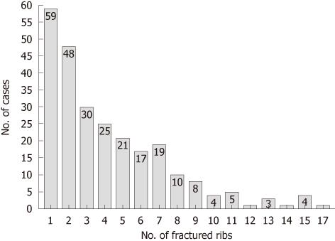
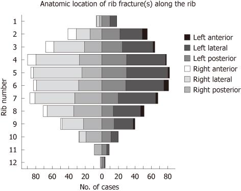
Of the 256 patients with rib fractures, 1074 ribs were fractured with a range of 1 to 17 fractures per patient (median number of fractured ribs = 3) (Figure 2). Of all ribs fractured, 107 were identified in patients with multiple fractures which consisted of 16 flail chests in 15 patients (5.9% of these 256 patients) (Figure 1B). The most common rib fractures were distributed from the 3rd through to the 8th ribs and the vast majority involved posterior and lateral locations (Figure 3). First and second rib fractures were observed in 84 patients (32.8% of these 256 patients).
Thoracic vertebral body fractures were detected in 60 patients (7.8%), 31 of which were involved in T1 through to T10 fractures (upper thoracic spine). Thirty-two of these patients also had rib fractures. Sternal fractures were detected in 2 patients. Fractures of the scapula and clavicle occurred in 63 patients (8.2%) and 45 of these patients also had rib fractures.
We found that rib fractures had a significant positive association with non-rib thoracic fractures (Table 1, P < 0.001). Furthermore, the number of fractured ribs was significantly higher in patients with non-rib thoracic fractures (Table 2, P < 0.001 according to the Wilcoxon W test in a comparison with patients without non-rib thoracic fractures).
| Rib fractures | Pearsonχ2 | P | ||
| + | - | |||
| Parenchymal injuries | 52 | 15 | 65.406 | < 0.001 |
| Pleural injuries | 113 | 22 | 188.573 | < 0.001 |
| Non-rib thoracic fractures | 67 | 48 | 38.441 | < 0.001 |
| No. of fractured ribs | Total | |||
| None | 1-2 | 3 or more | ||
| Non-rib thoracic fracture | ||||
| - | 468 | 80 | 109 | 657 |
| + | 48 | 27 | 40 | 115 |
| Total | 516 | 107 | 149 | 772 |
Sixty-seven patients had pulmonary contusion, 52 (77.6%) of which had coexisting rib fractures. Pleural injuries were noted in 135 patients, and 113 (83.7%) of these patients had coexisting rib fractures. Of these 135 patients, pleural complications included hemothorax (96, 71.1%), pneumothorax (15, 11.1%) and hemo-pneumothorax (24, 17.8%). Pleural drainage was performed in 24 cases (17.8%) with the laboratory results for blood detected in 21 patients (17 cases of hemothorax, four cases of hemo-pneumothorax).
Rib fractures had a significant positive association with pulmonary contusion and pleural injuries (Table 1, P < 0.001) and the incidence of coexisting pulmonary contusion and pleural injuries increased significantly in patients with 3 or more fractured ribs compared with patients with 1 or 2 fractured ribs (31.5% vs 4.7% for pulmonary contusion, and 56.4% vs 27.1% for pleural injuries).
Pneumomediastinum was found in 6 patients. All these patients had concurrent pleural injuries. Traumatic diaphragmatic hernia was detected in one patient on the left side. Subcutaneous air collection was detected in 29 patients and 25 of these patients had coexisting pleural injuries including 15 with pneumothorax or hemo-pneumothorax.
When the patients were divided into the two clinical outcome groups, fatal and discharged, logistic regression showed that pulmonary contusion (B = 2.27, χ2 = 10.401, P = 0.01) and the number of fractured ribs (χ2 = 12.739, P = 0.02) were significant factors associated with patient death in this cohort.
Chest trauma comprises 10%-15% of all traumas with a mortality rate ranging from 8.2% to 33.3%[10-13]. The leading cause of this type of trauma is road traffic accidents, and then falls, assault and work-related accidents[14]. However, the leading cause of thoracic injuries in the Sichuan earthquake was crush injuries, which were significantly different from other common thoracic traumas. We propose that the features of crush thoracic trauma associated with the earthquake were different from other common thoracic traumas.
Although there are some limitations in conventional screen-film radiography such as low image contrast, an AP chest radiograph remains the most common first imaging method used in patients with thoracic trauma[8,9]. Digital radiography can overcome the limitations of conventional screen-film radiography by significantly recovering the loss of image contrast with image processing, particularly with manipulation of image window width and window level[15,16]. We, therefore, focused on the assessment of crush thoracic trauma using CDR in 772 patients in the present study.
According to Sirmali et al[14], rib fractures are a common injury with an incidence of 38.7% in patients with common thoracic trauma. As shown in this study, the incidence of rib fractures was 33.2%, which was similar to their study as well as other studies on the Southern Hyogo Prefecture Earthquake (1995, Japan), Izmit region and Duzce region Earthquakes (1999, Turkey), and Bam earthquake (2003, Iran), where the incidence of rib fractures ranged from 27.0% to 36.4%[2-4]. The number of rib fractures is a good indicator of the severity of thoracic injuries. More than 3 rib fractures was associated with the greatest prognostic difference and the mortality rate increased with each additional rib fracture with total mortality ranging from 5.7% to 10%[12,14]. Fractures of the first and second rib after thoracic trauma indicate a severe injury, and the incidence of significant vascular injury can reach 14%[17]. In our series, multivariate logistic regression analysis also showed that the number of fractured ribs was a significant factor associated with patient death. Moreover, seven or more fractures occurred more frequently in our series than in two studies reported by Flagel et al[18] and Livingston et al[19] (21.9% vs 4.1% and 6.7%, respectively) which focused on common thoracic trauma. Furthermore, the mean number of rib fractures per patient, the incidence of fractures of the first and second rib and multiple fractures were also higher in our study than in previous studies on common thoracic traumas[12,14,18,19]. These findings may indicate that the crush injuries resulting from the earthquake were more severe than other common chest traumas. This may be because victims of the Sichuan earthquake might have fallen down in the prone or supine position, and were trapped. The horizontal position would have made the victims more vulnerable to persistent bilateral compression generated by falling objects, and was strikingly different from the kinetic energy of other common chest traumas.
The thoracic spine is much stiffer than the lumbar spine in sagittal and lateral flexion-extension. Fractures in the upper thoracic spine (T1-T10) comprised only 16% of the thoracic spine fractures[20]. The incidence of fractures of the sternum, scapulae and clavicles was low, and a large force is required to break these bones. A sternal fracture could be interpreted as a marker lesion for severe trauma[21].
As shown in this study, a high incidence of upper thoracic vertebral fractures (51.7%) was detected. This could be explained by the random crush energy exerted on the spine which might have been more frequent than falls and other common injuries, and may show that the injuries resulting from the earthquake were widespread multi-regional injuries. We also noted that rib fractures had a significant positive association with non-rib thoracic fractures. The number of fractured ribs was significantly higher in patients with non-rib thoracic fractures. Thus, the relatively high incidence of these non-rib thoracic fractures in our patients could be more evidence of the severity of earthquake trauma.
As major complications of rib fractures, lung contusions, pneumothorax and hemothorax have more clinical impact than the fracture itself. Pulmonary contusion is the most common thoracic injury with a morbidity ranging from 23.7% to 49.6%[11,12,18]. It can develop rapidly within the first 6 h after injury, and was seen as an ill-defined area of air space opacity on CDR that was not impeded by the pleural fissure, and would heal with little residual scarring within 7-14 d. Pleural injuries are the second most common traumatic conditions of chest trauma with an incidence of 41.8%, and were noted in 72.3% of cases with rib fractures[12,14]. Although hemothorax was not a clinically significant indicator of poor prognosis, ongoing accumulation of blood may require tube thoracostomy or open thoracotomy; a tension pneumothorax is a clinical emergency and should be considered when the patient’s condition rapidly deteriorates[12].
The incidence of pulmonary contusion and pleural injuries in this study was similar with that in the other two studies on earthquake injuries[2,3]. The significance of pulmonary contusion as a significant factor associated with patient death was revealed by multivariate logistic regression analysis. Furthermore, a positive association between pulmonary contusion and rib fractures, as well as that between pleural injuries and rib fractures was found in this series. As mentioned above, rib fractures had a significant positive association with non-rib thoracic fractures. Therefore, abnormalities of the bony thorax including ribs, clavicles, scapulae, and spine noted on a supine AP CDR might suggest severe injury that is likely to involve other thoracic structures[8].
A very low incidence of pneumomediastinum was detected in this study. This was perhaps due to the high proportion of supine AP CDR and the relatively lower image contrast of CDR. Furthermore, the late transfer time to hospital may be another reason. The mean time from injury to CDR was 6 d, which was due to the disaster zones of this massive earthquake being mainly mountain areas with severe road destruction, and insufficient rescue as some of the hospitals in the disaster zone were damaged with severe staff casualties. This may have been similar to the Taiwan (1999) earthquake, in which the long transportation times to back-up hospitals due to impassable roads and disrupted communication systems were the most important reasons for inadequate response and resuscitation of severely injured victims[22].
Traumatic diaphragmatic rupture accounts for 4% to 8% of all organ injuries from blunt trauma including tears, ruptures, avulsions, and eventually late necrosis[23,24]. In adults, left sided traumatic diaphragmatic rupture is predominant[25]. We had a very low incidence of patients diagnosed with diaphragmatic rupture, as this condition may have been present with other severe abdominal organ injuries and the victims died before they were rescued.
Our study has some limitations. First, as noted in our cohort, more than half of these patients received only an AP CDR and almost 90% underwent supine AP CDR. This may have made the evaluation of some types of injuries difficult and insufficient. However, as a starting point in the imaging work-up, CDR revealed the severity of crush thoracic trauma by detecting thoracic cage fractures. This was important for the primary diagnostic work-up, and for the choice of adjunctive imaging method, computed tomography. Second, because the soft tissue injuries shown on CDR were ambiguous, the diagnosis of these injuries such as edema, contusion and laceration may have been ignored in this study, which may decrease the morbidity of thoracic injuries in this cohort. However it did not influence the evaluation of severity.
We conclude that crush thoracic trauma in the massive Sichuan earthquake resulted in severe thoracic cage fractures, which was perhaps due to the special mechanism of this type of injury. Rib fractures often coexisted with non-rib thoracic fractures, pulmonary contusion and pleural injuries. CDR revealed the severity of these life threatening traumas. We strongly stress the high number of rib fractures and pulmonary contusions in this study which were the most severe injuries and needed appropriate medical treatment, because these were significant factors associated with patient death.
An earthquake with a magnitude of 8.0 occurred at 2:28 PM on May 12, 2008 in Sichuan province and injured thousands of people. Most were crushed due to building collapse, flying stones, bricks or other falling objects. As the injured must be retrieved from beneath collapsed rubble, earthquake-related thoracic injuries still present a major challenge to emergency medical staff. An antero-posterior (AP) chest radiograph remains the most common first imaging method obtained in patients with thoracic trauma. Thus, the authors retrospectively reviewed the chest digital radiography (CDR) appearance of 772 consecutive patients injured in the massive Sichuan earthquake to investigate the features of crush thoracic trauma.
This article focused on earthquake-related crush thoracic trauma patients injured in the 2008 massive earthquake. We cite better disaster preparedness as an overarching goal of this investigation.
The CDR characteristics of crush thoracic trauma in Sichuan earthquake victims were described. We believe that CDR can reveal the severity of this life threatening trauma. The high number of rib fractures and pulmonary contusions were significant prognostic factors which needed appropriate medical treatment.
As a starting point in the imaging work-up, we found that CDR revealed the severity of crush thoracic trauma by detecting thoracic cage fractures. This may be important for the primary diagnostic work-up, and for the choice of adjunctive imaging method, computed tomography. However, as more than half of these patients received only an AP CDR and almost 90% underwent supine AP CDR, the evaluation of some types of injuries such as patients with hemothorax and/or pneumothorax, pulmonary parenchymal injuries and occult bony fractures was difficult.
This is a well understood article. A detailed description in imaging characteristics of crush thoracic trauma in Sichuan earthquake is provided. The sample size is adequate. The statistical methods used are appropriate.
Peer reviewers: Edson Marchiori, MD, PhD, Full Professor of Radiology, Department of Radiology, Fluminense Federal University, Rua Thomaz Cameron, 438, Valparaiso, CEP 25685, 120, Petrópolis, Rio de Janeiro, Brazil; George Panayiotakis, Professor, Department of Medical Physics, School of MedicineUniversity of Patras, 26500, Patras, Greece
S- Editor Cheng JX L- Editor Webster JR E- Editor Zheng XM
| 1. | Dong ZH, Yang ZG, Chen TW, Feng YC, Wang QL, Chu ZG. Spinal injuries in the Sichuan earthquake. N Engl J Med. 2009;361:636-637. [RCA] [PubMed] [DOI] [Full Text] [Cited by in Crossref: 13] [Cited by in RCA: 51] [Article Influence: 3.2] [Reference Citation Analysis (0)] |
| 2. | Yoshimura N, Nakayama S, Nakagiri K, Azami T, Ataka K, Ishii N. Profile of chest injuries arising from the 1995 southern Hyogo Prefecture earthquake. Chest. 1996;110:759-761. [PubMed] |
| 3. | Ozdoğan S, Hocaoğlu A, Cağlayan B, Imamoğlu OU, Aydin D. Thorax and lung injuries arising from the two earthquakes in Turkey in 1999. Chest. 2001;120:1163-1166. [PubMed] |
| 4. | Ghodsi SM, Zargar M, Khaji A, Karbakhsh M. Chest injury in victims of Bam earthquake. Chin J Traumatol. 2006;9:345-348. [PubMed] |
| 5. | Toker A, Isitmangil T, Erdik O, Sancakli I, Sebit S. Analysis of chest injuries sustained during the 1999 Marmara earthquake. Surg Today. 2002;32:769-771. [RCA] [PubMed] [DOI] [Full Text] [Cited by in Crossref: 10] [Cited by in RCA: 13] [Article Influence: 0.6] [Reference Citation Analysis (0)] |
| 6. | Gautschi OP, Cadosch D, Rajan G, Zellweger R. Earthquakes and trauma: review of triage and injury-specific, immediate care. Prehosp Disaster Med. 2008;23:195-201. [PubMed] |
| 7. | Dong ZH, Yang ZG, Chen TW, Feng YC, Chu ZG, Yu JQ, Bai HL, Wang QL. Crush thoracic trauma in the massive Sichuan earthquake: evaluation with multidetector CT of 215 cases. Radiology. 2010;254:285-291. [PubMed] |
| 8. | Carpenter AJ. Diagnostic techniques in thoracic trauma. Semin Thorac Cardiovasc Surg. 2008;20:2-5. [RCA] [PubMed] [DOI] [Full Text] [Cited by in Crossref: 3] [Cited by in RCA: 4] [Article Influence: 0.2] [Reference Citation Analysis (0)] |
| 9. | Flores HA, Stewart RM. The multiply injured patient. Semin Thorac Cardiovasc Surg. 2008;20:64-68. [RCA] [PubMed] [DOI] [Full Text] [Cited by in Crossref: 2] [Cited by in RCA: 3] [Article Influence: 0.2] [Reference Citation Analysis (0)] |
| 10. | Ziegler DW, Agarwal NN. The morbidity and mortality of rib fractures. J Trauma. 1994;37:975-979. [RCA] [PubMed] [DOI] [Full Text] [Cited by in Crossref: 408] [Cited by in RCA: 454] [Article Influence: 14.6] [Reference Citation Analysis (0)] |
| 11. | Balci AE, Kazez A, Eren S, Ayan E, Ozalp K, Eren MN. Blunt thoracic trauma in children: review of 137 cases. Eur J Cardiothorac Surg. 2004;26:387-392. [RCA] [PubMed] [DOI] [Full Text] [Cited by in Crossref: 78] [Cited by in RCA: 66] [Article Influence: 3.1] [Reference Citation Analysis (0)] |
| 12. | Freixinet J, Beltrán J, Rodríguez PM, Juliá G, Hussein M, Gil R, Herrero J. [Indicators of severity in chest trauma]. Arch Bronconeumol. 2008;44:257-262. [PubMed] |
| 13. | Virgós Señor B, Nebra Puertas AC, Sánchez Polo C, Broto Civera A, Suárez Pinilla MA. [Predictors of outcome in blunt chest trauma]. Arch Bronconeumol. 2004;40:489-494. [RCA] [PubMed] [DOI] [Full Text] [Cited by in Crossref: 6] [Cited by in RCA: 6] [Article Influence: 0.3] [Reference Citation Analysis (0)] |
| 14. | Sirmali M, Türüt H, Topçu S, Gülhan E, Yazici U, Kaya S, Taştepe I. A comprehensive analysis of traumatic rib fractures: morbidity, mortality and management. Eur J Cardiothorac Surg. 2003;24:133-138. [RCA] [PubMed] [DOI] [Full Text] [Cited by in Crossref: 324] [Cited by in RCA: 335] [Article Influence: 15.2] [Reference Citation Analysis (0)] |
| 15. | Behrman RH, Yasuda G. Effective dose in diagnostic radiology as a function of x-ray beam filtration for a constant exit dose and constant film density. Med Phys. 1998;25:780-790. [RCA] [PubMed] [DOI] [Full Text] [Cited by in Crossref: 18] [Cited by in RCA: 19] [Article Influence: 0.7] [Reference Citation Analysis (0)] |
| 16. | Hamer OW, Sirlin CB, Strotzer M, Borisch I, Zorger N, Feuerbach S, Völk M. Chest radiography with a flat-panel detector: image quality with dose reduction after copper filtration. Radiology. 2005;237:691-700. [PubMed] |
| 17. | Livoni JP, Barcia TC. Fracture of the first and second rib: incidence of vascular injury relative to type of fracture. Radiology. 1982;145:31-33. [PubMed] |
| 18. | Flagel BT, Luchette FA, Reed RL, Esposito TJ, Davis KA, Santaniello JM, Gamelli RL. Half-a-dozen ribs: the breakpoint for mortality. Surgery. 2005;138:717-723; discussion 723-725. [RCA] [PubMed] [DOI] [Full Text] [Cited by in Crossref: 296] [Cited by in RCA: 344] [Article Influence: 17.2] [Reference Citation Analysis (0)] |
| 19. | Livingston DH, Shogan B, John P, Lavery RF. CT diagnosis of Rib fractures and the prediction of acute respiratory failure. J Trauma. 2008;64:905-911. [RCA] [PubMed] [DOI] [Full Text] [Cited by in Crossref: 99] [Cited by in RCA: 100] [Article Influence: 5.9] [Reference Citation Analysis (0)] |
| 20. | Meyer PR. Fractures of the thoracic spine: T1 to T10. Surgery of spine trauma. New York: Churchill Livingstone 2007; 527-571. |
| 21. | Knobloch K, Wagner S, Haasper C, Probst C, Krettek C, Vogt PM, Otte D, Richter M. Sternal fractures are frequent among polytraumatised patients following high deceleration velocities in a severe vehicle crash. Injury. 2008;39:36-43. [RCA] [PubMed] [DOI] [Full Text] [Cited by in Crossref: 11] [Cited by in RCA: 12] [Article Influence: 0.7] [Reference Citation Analysis (0)] |
| 22. | Yi-Szu W, Chung-Ping H, Tzu-Chieh L, Dar-Yu Y, Tain-Cheng W. Chest injuries transferred to trauma centers after the 1999 Taiwan earthquake. Am J Emerg Med. 2000;18:825-827. [RCA] [PubMed] [DOI] [Full Text] [Cited by in Crossref: 13] [Cited by in RCA: 15] [Article Influence: 0.6] [Reference Citation Analysis (0)] |
| 23. | Cupitt JM, Smith MB. Missed diaphragm rupture following blunt trauma. Anaesth Intensive Care. 2001;29:292-296. [PubMed] |
| 24. | Seleem MI, Al-Hashemy AM. Delayed presentation of traumatic rupture of the diaphragm. Saudi Med J. 2001;22:714-717. [PubMed] |
| 25. | Esme H, Solak O, Sahin DA, Sezer M. Blunt and penetrating traumatic ruptures of the diaphragm. Thorac Cardiovasc Surg. 2006;54:324-327. [RCA] [PubMed] [DOI] [Full Text] [Cited by in Crossref: 24] [Cited by in RCA: 20] [Article Influence: 1.1] [Reference Citation Analysis (0)] |


