Published online Jul 28, 2021. doi: 10.4329/wjr.v13.i7.233
Peer-review started: January 29, 2021
First decision: March 17, 2021
Revised: March 30, 2021
Accepted: June 15, 2021
Article in press: June 15, 2021
Published online: July 28, 2021
Processing time: 173 Days and 7.1 Hours
In chest computed tomography (CT) scan, bilateral peripheral multifocal ground-glass opacities, linear opacities, reversed halo sign, and crazy-paving pattern are suggestive for coronavirus disease 2019 (COVID-19) in clinically suspicious cases, but they are not specific for the diagnosis, as other viral pneumonias, like influ
To find a specific imaging feature of the disease would be a welcome guide in diagnosis and management of challenging cases.
Chest CT imaging findings of 650 patients admitted to a university Hospital in Tehran, Iran between January 2020 and July 2020 with confirmed COVID-19 in
PTS were presented in 32 cases (frequency 4.9%). The location of the lesions in 31 of the 32 cases (96.8%) was peripheral, while 4 of the 31 cases had lesions both peripherally and centrally. In 25 cases, the lesions were located near the pleural surface and considered pleural based and half of the lesions (at least one lesion) were in the lower segments and lobes of the lungs. 22 cases had multiple lesions with a > 68% frequency. More than 87% of cases had an adjacent bronchovascular bundle. Ground-glass opacities were detectable adjacent or close to the lesions in 30 cases (93%) and only in 7 cases (21%) was consolidation adjacent to the lesions.
Although it is not frequent in COVID-19, familiarity with this feature may help radiologists and physicians distinguish the disease from other viral and non-infectious pneumonias in challenging cases.
Core Tip: In this report, a new diagnostic imaging sign in chest computed tomography of coronavirus disease 2019 cases, the “pulmonary target sign”, is reported and its characteristics are described. Previous reports are limited to a small number of case reports and this appearance is not fully described.
- Citation: Jafari R, Jonaidi-Jafari N, Maghsoudi H, Dehghanpoor F, Schoepf UJ, Ulversoy KA, Saburi A. “Pulmonary target sign” as a diagnostic feature in chest computed tomography of COVID-19. World J Radiol 2021; 13(7): 233-242
- URL: https://www.wjgnet.com/1949-8470/full/v13/i7/233.htm
- DOI: https://dx.doi.org/10.4329/wjr.v13.i7.233
Coronavirus disease 2019 (COVID-19) is the seventh member of the non-segmented, enveloped, and positive-sense-RNA Coronaviridae family, which causes acute respiratory illness. This new coronavirus was first detected in Wuhan, China, in December 2019. It has since rapidly spread throughout the world and was recognized as a global health emergency[1,2]. COVID-19 presents as a wide spectrum of clinical pictures, from asymptomatic or mild flu-like illness to severe respiratory infection and even death[3,4].
A definitive diagnosis of COVID-19 mainly relies on RT-PCR testing in suspected cases. Chest computed tomography (CT) also has an undeniable importance in the diagnostic management of COVID-19 due to its high sensitivity and widespread availability[5]. The most common radiologic findings of COVID-19 are bilateral, peripheral, multifocal ground-glass opacities (GGO) and consolidations, linear opa
Some relatively specific features of the disease in chest CT have been discussed in the literature, including the “parallel pleural sign”, “rings of Saturn appearance” and, recently, the “pulmonary target sign (PTS)”[9,10]. The latter imaging finding seems to be more specific for the disease. It was initially reported by Jafari et al[11] and Shaghaghi et al[12] as a hyperattenuating ring surrounding a dense central dot, mi
Chest CT imaging findings of 650 patients admitted to a university Hospital in Tehran, Iran with confirmed COVID-19 infection by RT-PCR between January 2020 and July 2020 were reviewed by two expert radiologists.
All chest CT scan were obtained using a 16-row detector CT scanner (GE, optima, United States). Based on protocol of COVID-19 low-dose thoracic CT scan, the fo
In addition to common non-specific imaging findings of COVID-19 pneumonia, radiologic characteristics of PTS will be presented. This chest CT sign of the disease as a circular appearance of non-involved pulmonary parenchyma with a central hyper
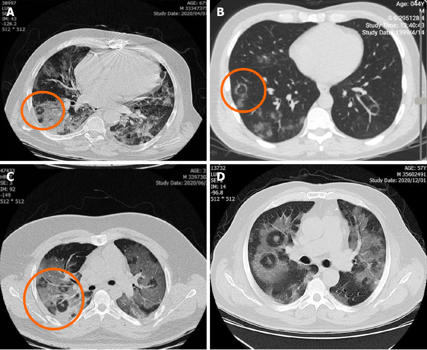
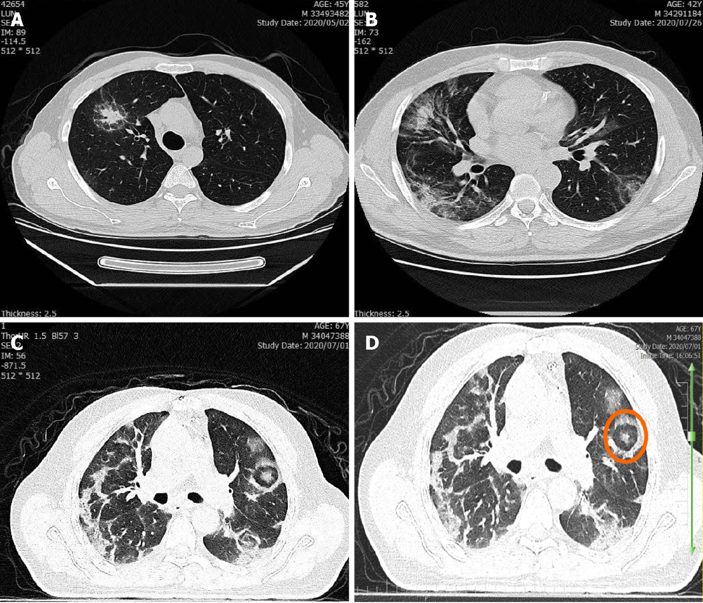
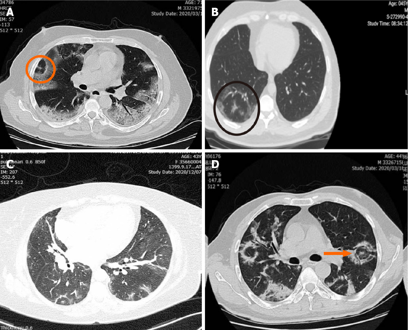
Of the 650 patients reviewed, 32 cases of PTS were found (4.9% prevalence). The location of the lesions in 31 of the 32 cases was peripheral, while 4 of the 31 cases had lesions both peripherally and centrally. Only one case had an isolated central lesion mimicking a solitary pulmonary nodule (Figures 1 and 2A).
The typical shape of PTS was seen in 31 cases, while 1 case had a PTS variant with double peripheral dense rings, which was previously named “rings of Saturn” (see Figure 2).
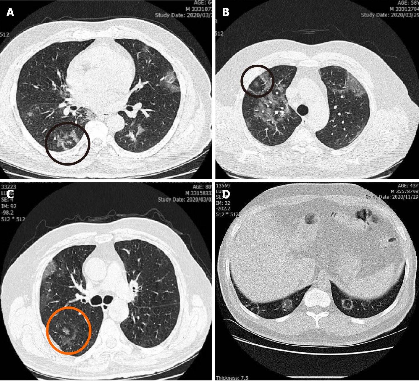
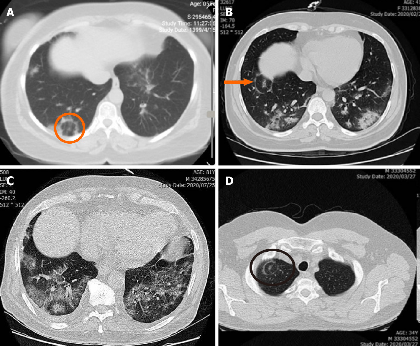
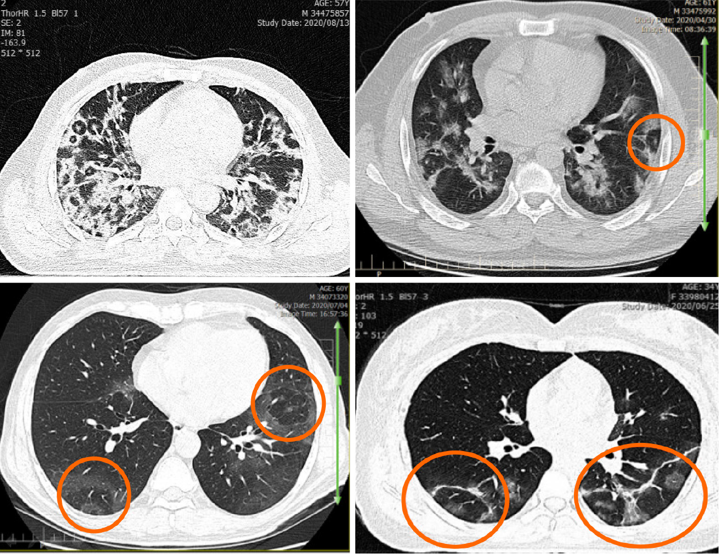
In 25 cases, the lesions (at least one if there were multiple) were located near the pleural surface and considered pleural based (see Figure 3). Half of the lesions (at least one lesion) were in the lower segments and lobes of the lungs (see Figure 4).
More than 87% of cases had an adjacent bronchovascular bundle (BVB). This characteristic was reported when a dense branching linear structure was approaching the lesion (see Figure 5).
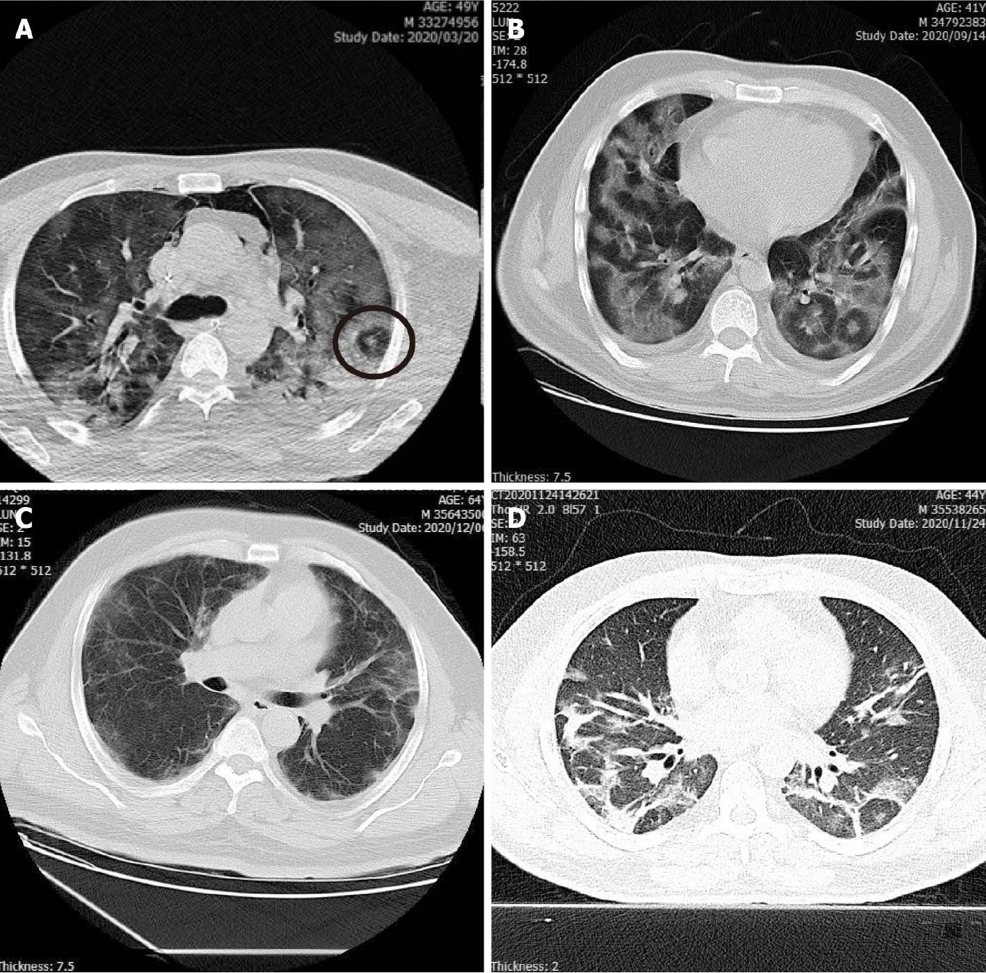
Of the 32 cases, 22 had multiple lesions with a > 68% frequency (see Figure 6). GGOs were detectable adjacent or close to the lesions in 30 cases (93%) and only in 7 cases (21%) was consolidation adjacent to the lesions, Figure 7.
8 cases showed pulmonary complications of COVID-19, including pneumothorax (1 case) and pleural effusion (7 cases/21%). Three cases (9%) showed parallel pleural sign and 6 cases (18%) showed fibrotic bands (see Figure 8). The characteristics are summarized in Table 1.
Regarding the descriptive findings and characteristics of PTS lesions, they tend to be multiple lesions, located in the periphery, and located adjacent to a BVB and GGOs. They are uncommonly seen centrally or basally or with adjoining consolidation. Due to a low frequency of fibrotic bands as a marker of healing and concomitant complications, such as pleural effusion, it seems that PTS appear at early phases.
In such contagious and life-threatening infections as COVID-19, having a consistent and reliable diagnostic and screening tool is vital. Currently, CT, with its high sensitivity and specificity, is one of the most valuable screening and diagnostic tools[17,18]. Although commonly reported findings in COVID-19 CT scans are not specific for a diagnosis of COVID-19 vs other viral pneumonias, some recently reported specific features of the disease, like PTS, can be helpful for this aim.
It is important to know the difference between PTS and the Atoll sign. An Atoll sign has central opacities consisting of GGO, while PTS has a central dot which can represent a filled bronchiole or vessel. Moreover, it was previously noted that “the crescentic appearance of the reversed halo sign is typical on CT whereas the target sign has a polygonal appearance peripherally”[19]. This feature has been frequently re
Generally, diffuse subpleural and peripheral ill-defined GGO with air-broncho
In our contribution, we present 32 PCR confirmed cases of COVID-19 infection with specific findings on their chest CT. As mentioned previously, in addition to common findings of COVID-19 infection, their chest CT revealed a circular appearance of non-involved pulmonary parenchyma, which encompassed a central hyperdense dot surrounded by ground-glass or alveolar opacities. This represents a unique finding that has never been reported in any other disease. We hypothesize that this appea
We present specific, unique chest CT imaging features in 32 confirmed cases of COVID-19 infection. Although these findings are not observed in all patients with this disease and it is uncommon (about 5% frequency), we believe PTS to be a specific finding which can distinguish COVID-19 pneumonia from other similar viral pneu
Chest computed tomography scan findings like bilateral ground glass opacities and consolidations are commonly used as distinguishing features in the differential diagnosis of coronavirus disease 2019 (COVID-19). However, a problem in diagnosis arises when other viral or atypical pneumonia infections are suspected, as they may present similarly.
Pulmonary target sign (PTS) is a feature of COVID-19 that has been recently suggested as an atypical presentation of pulmonary involvement and may be used to distinguish COVID-19 from other similar pneumonia infections.
In this paper, the PTS and its characteristics were assessed among COVID-19 confirm
Among all cases of COVID-19 that were referred to a tertiary medical center in Tehran, Iran, chest CT scan findings of 650 serologically positive cases of COVID-19 were evaluated for PTS and its characteristics.
32 individuals with at least one PTS in their CT scan were identified in which most of the PTSs were multiple in number, in a peripheral location, and near a bronchovas
The PTS has a frequency of about 5% and specific characteristics that may make it useful in the prompt diagnosis of COVID-19.
The relationship between the presence of the PTS and the prognosis of COVID-19 still needs to be elucidated. Additionally, the mechanisms behind the pathogenesis and the timeline of PTS progression are suggested areas of research for future studies.
Manuscript source: Invited manuscript
Specialty type: Radiology, nuclear medicine and medical imaging
Country/Territory of origin: Iran
Peer-review report’s scientific quality classification
Grade A (Excellent): 0
Grade B (Very good): 0
Grade C (Good): C
Grade D (Fair): 0
Grade E (Poor): 0
P-Reviewer: Chen X S-Editor: Liu M L-Editor: Filipodia P-Editor: Yuan YY
| 1. | Wang D, Hu B, Hu C, Zhu F, Liu X, Zhang J, Wang B, Xiang H, Cheng Z, Xiong Y, Zhao Y, Li Y, Wang X, Peng Z. Clinical Characteristics of 138 Hospitalized Patients With 2019 Novel Coronavirus-Infected Pneumonia in Wuhan, China. JAMA. 2020;323:1061-1069. [RCA] [PubMed] [DOI] [Full Text] [Cited by in Crossref: 14113] [Cited by in RCA: 14764] [Article Influence: 2952.8] [Reference Citation Analysis (0)] |
| 2. | Shi H, Han X, Jiang N, Cao Y, Alwalid O, Gu J, Fan Y, Zheng C. Radiological findings from 81 patients with COVID-19 pneumonia in Wuhan, China: a descriptive study. Lancet Infect Dis. 2020;20:425-434. [RCA] [PubMed] [DOI] [Full Text] [Full Text (PDF)] [Cited by in Crossref: 2493] [Cited by in RCA: 2310] [Article Influence: 462.0] [Reference Citation Analysis (0)] |
| 3. | Kanne JP, Little BP, Chung JH, Elicker BM, Ketai LH. Essentials for Radiologists on COVID-19: An Update-Radiology Scientific Expert Panel. Radiology. 2020;296:E113-E114. [RCA] [PubMed] [DOI] [Full Text] [Full Text (PDF)] [Cited by in Crossref: 367] [Cited by in RCA: 421] [Article Influence: 84.2] [Reference Citation Analysis (0)] |
| 4. | Saburi A, Schoepf UJ, Ulversoy KA, Jafari R, Eghbal F, Ghanei M. From Radiological Manifestations to Pulmonary Pathogenesis of COVID-19: A Bench to Bedside Review. Radiol Res Pract. 2020;2020:8825761. [RCA] [PubMed] [DOI] [Full Text] [Full Text (PDF)] [Cited by in Crossref: 6] [Cited by in RCA: 6] [Article Influence: 1.2] [Reference Citation Analysis (0)] |
| 5. | Li M, Lei P, Zeng B, Li Z, Yu P, Fan B, Wang C, Zhou J, Hu S, Liu H. Coronavirus Disease (COVID-19): Spectrum of CT Findings and Temporal Progression of the Disease. Acad Radiol. 2020;27:603-608. [RCA] [PubMed] [DOI] [Full Text] [Full Text (PDF)] [Cited by in Crossref: 192] [Cited by in RCA: 170] [Article Influence: 34.0] [Reference Citation Analysis (0)] |
| 6. | Simpson S, Kay FU, Abbara S, Bhalla S, Chung JH, Chung M, Henry TS, Kanne JP, Kligerman S, Ko JP, Litt H. Radiological Society of North America Expert Consensus Statement on Reporting Chest CT Findings Related to COVID-19. Endorsed by the Society of Thoracic Radiology, the American College of Radiology, and RSNA - Secondary Publication. J Thorac Imaging. 2020;35:219-227. [RCA] [PubMed] [DOI] [Full Text] [Full Text (PDF)] [Cited by in Crossref: 400] [Cited by in RCA: 564] [Article Influence: 112.8] [Reference Citation Analysis (0)] |
| 7. | Hosseiny M, Kooraki S, Gholamrezanezhad A, Reddy S, Myers L. Radiology Perspective of Coronavirus Disease 2019 (COVID-19): Lessons From Severe Acute Respiratory Syndrome and Middle East Respiratory Syndrome. AJR Am J Roentgenol. 2020;214:1078-1082. [RCA] [PubMed] [DOI] [Full Text] [Cited by in Crossref: 257] [Cited by in RCA: 266] [Article Influence: 53.2] [Reference Citation Analysis (0)] |
| 8. | Hanfi SH, Lalani TK, Saghir A, McIntosh LJ, Lo HS, Kotecha HM. COVID-19 and its Mimics: What the Radiologist Needs to Know. J Thorac Imaging. 2021;36:W1-W10. [RCA] [PubMed] [DOI] [Full Text] [Cited by in Crossref: 17] [Cited by in RCA: 17] [Article Influence: 4.3] [Reference Citation Analysis (0)] |
| 9. | Wang C, Shi B, Wei C, Ding H, Gu J, Dong J. Initial CT features and dynamic evolution of early-stage patients with COVID-19. Radiol Infect Dis. 2020;7:195-203. [RCA] [PubMed] [DOI] [Full Text] [Full Text (PDF)] [Cited by in Crossref: 6] [Cited by in RCA: 9] [Article Influence: 1.8] [Reference Citation Analysis (0)] |
| 10. | Wu J, Pan J, Teng D, Xu X, Feng J, Chen YC. Interpretation of CT signs of 2019 novel coronavirus (COVID-19) pneumonia. Eur Radiol. 2020;30:5455-5462. [RCA] [PubMed] [DOI] [Full Text] [Full Text (PDF)] [Cited by in Crossref: 46] [Cited by in RCA: 62] [Article Influence: 12.4] [Reference Citation Analysis (0)] |
| 11. | Jafari R, M Colletti P, Saburi A. "Rings of Saturn" appearance: a unique finding in a case of COVID-19 pneumonitis. Diagn Interv Radiol. 2021;27:154. [RCA] [PubMed] [DOI] [Full Text] [Cited by in Crossref: 7] [Cited by in RCA: 4] [Article Influence: 1.0] [Reference Citation Analysis (0)] |
| 12. | Shaghaghi S, Daskareh M, Irannejad M, Shaghaghi M, Kamel IR. Target-shaped combined halo and reversed-halo sign, an atypical chest CT finding in COVID-19. Clin Imaging. 2021;69:72-74. [RCA] [PubMed] [DOI] [Full Text] [Full Text (PDF)] [Cited by in Crossref: 18] [Cited by in RCA: 20] [Article Influence: 4.0] [Reference Citation Analysis (0)] |
| 13. | McLaren TA, Gruden JF, Green DB. The bullseye sign: A variant of the reverse halo sign in COVID-19 pneumonia. Clin Imaging. 2020;68:191-196. [RCA] [PubMed] [DOI] [Full Text] [Full Text (PDF)] [Cited by in Crossref: 25] [Cited by in RCA: 17] [Article Influence: 3.4] [Reference Citation Analysis (0)] |
| 14. | Gomes de Farias LP, Caixeta Souza FH, da Silva Teles GB. The Target Sign and Its Variant in COVID-19 Pneumonia. Radiol Cardiothorac Imaging. 2020;2:e200435. [RCA] [PubMed] [DOI] [Full Text] [Full Text (PDF)] [Cited by in Crossref: 8] [Cited by in RCA: 6] [Article Influence: 1.2] [Reference Citation Analysis (0)] |
| 15. | Müller CIS, Müller NL. Chest CT target sign in a couple with COVID-19 pneumonia. Radiol Bras. 2020;53:252-254. [RCA] [PubMed] [DOI] [Full Text] [Full Text (PDF)] [Cited by in Crossref: 27] [Cited by in RCA: 24] [Article Influence: 4.8] [Reference Citation Analysis (0)] |
| 16. | Jafari R, Maghsoudi H, Saburi A. A Unique Feature of COVID-19 Infection in Chest CT; "Pulmonary Target" Appearance. Acad Radiol. 2021;28:146-147. [RCA] [PubMed] [DOI] [Full Text] [Full Text (PDF)] [Cited by in Crossref: 8] [Cited by in RCA: 5] [Article Influence: 1.3] [Reference Citation Analysis (0)] |
| 17. | Ai T, Yang Z, Hou H, Zhan C, Chen C, Lv W, Tao Q, Sun Z, Xia L. Correlation of Chest CT and RT-PCR Testing for Coronavirus Disease 2019 (COVID-19) in China: A Report of 1014 Cases. Radiology. 2020;296:E32-E40. [RCA] [PubMed] [DOI] [Full Text] [Full Text (PDF)] [Cited by in Crossref: 3614] [Cited by in RCA: 3284] [Article Influence: 656.8] [Reference Citation Analysis (0)] |
| 18. | Goyal N, Chung M, Bernheim A, Keir G, Mei X, Huang M, Li S, Kanne JP. Computed Tomography Features of Coronavirus Disease 2019 (COVID-19): A Review for Radiologists. J Thorac Imaging. 2020;35:211-218. [RCA] [PubMed] [DOI] [Full Text] [Cited by in Crossref: 23] [Cited by in RCA: 23] [Article Influence: 4.6] [Reference Citation Analysis (0)] |
| 19. | Marchiori E, Nobre LF, Hochhegger B, Zanetti G. CT characteristics of COVID-19: reversed halo sign or target sign? Diagn Interv Radiol. 2021;27:306-307. [RCA] [PubMed] [DOI] [Full Text] [Cited by in Crossref: 1] [Cited by in RCA: 3] [Article Influence: 0.8] [Reference Citation Analysis (0)] |
| 20. | Hoda A, Arash S. The Role of Chest CT Scan in Diagnosis of COVID-19. Adv J Emerg Med. 2020;4:e64. [DOI] [Full Text] |
| 21. | hao L, Chen B, Huang Y, Yang M, Yang J, Zhao Z. A Dynamic Follow-Up of Pneumonia Caused by Coronavirus Disease 2019 (COVID-19) on CT Scan. Iran J Radiol. 2020;17:e102847. [RCA] [DOI] [Full Text] [Cited by in Crossref: 2] [Cited by in RCA: 3] [Article Influence: 0.6] [Reference Citation Analysis (0)] |
| 22. | Zhang FY, Qiao Y, Zhang H. CT imaging of the COVID-19. J Formos Med Assoc. 2020;119:990-992. [RCA] [PubMed] [DOI] [Full Text] [Full Text (PDF)] [Cited by in Crossref: 10] [Cited by in RCA: 11] [Article Influence: 2.2] [Reference Citation Analysis (0)] |
| 23. | Kambouchner M, Bernaudin JF. Intralobular pulmonary lymphatic distribution in normal human lung using D2-40 antipodoplanin immunostaining. J Histochem Cytochem. 2009;57:643-648. [RCA] [PubMed] [DOI] [Full Text] [Cited by in Crossref: 44] [Cited by in RCA: 45] [Article Influence: 2.8] [Reference Citation Analysis (0)] |
| 24. | Elsoukkary SS, Mostyka M, Dillard A, Berman DR, Ma LX, Chadburn A, Yantiss RK, Jessurun J, Seshan SV, Borczuk AC, Salvatore SP. Autopsy Findings in 32 Patients with COVID-19: A Single-Institution Experience. Pathobiology. 2021;88:56-68. [RCA] [PubMed] [DOI] [Full Text] [Full Text (PDF)] [Cited by in Crossref: 66] [Cited by in RCA: 97] [Article Influence: 19.4] [Reference Citation Analysis (0)] |


