Copyright
©The Author(s) 2017.
World J Radiol. Jun 28, 2017; 9(6): 280-286
Published online Jun 28, 2017. doi: 10.4329/wjr.v9.i6.280
Published online Jun 28, 2017. doi: 10.4329/wjr.v9.i6.280
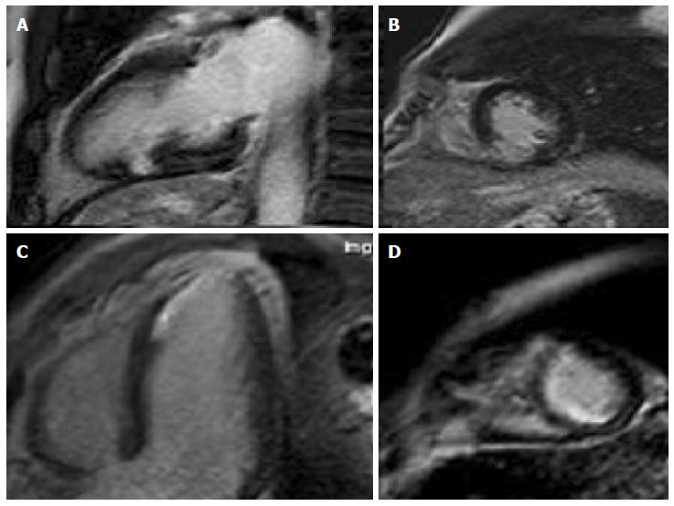
Figure 1 Cardiac magnetic resonance in myocardial infarction: Areas of subendocardial late gadolinium enhancement.
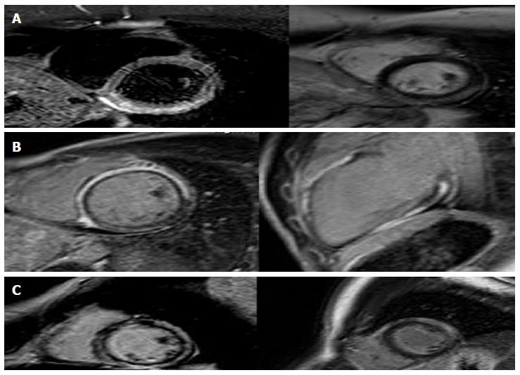
Figure 2 Cardiac magnetic resonance in myocarditis: (A) area of subepicardial edema (B-C) post contrast images show enhancement in the same area.
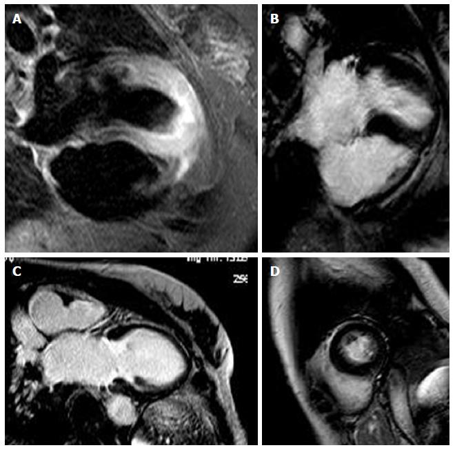
Figure 3 Cardiac magnetic resonance in apical ballooning: T2 images show edema in the apical segments (A), cine-MR shows akinesia (B: Diastole; C: Systole) in the same segments without (D) late gadolinium enhancement in the postcontrast images.
- Citation: Camastra GS, Sbarbati S, Danti M, Cacciotti L, Semeraro R, Della Sala SW, Ansalone G. Cardiac magnetic resonance in patients with acute cardiac injury and unobstructed coronary arteries. World J Radiol 2017; 9(6): 280-286
- URL: https://www.wjgnet.com/1949-8470/full/v9/i6/280.htm
- DOI: https://dx.doi.org/10.4329/wjr.v9.i6.280


