Copyright
©The Author(s) 2017.
World J Radiol. May 28, 2017; 9(5): 245-252
Published online May 28, 2017. doi: 10.4329/wjr.v9.i5.245
Published online May 28, 2017. doi: 10.4329/wjr.v9.i5.245
Figure 1 Patient with evidence of a ca.
32 mm tumor with arterial blood flow typical of hepatocellular carcinoma (wash-in phase at the centre of the image) in segment 3 treated with a single cycle of transarterial chemoembolization. A: Corresponding histological image of the tumor after transplantation: Particles can be seen inside the afferent arterioles of the tumor, documenting 100% necrosis; B: The follow-up CT scan after TACE observed complete response according to mRECIST. TACE: Transarterial chemoembolization; mRECIST: Modified Response Evaluation Criteria in Solid Tumors; CT: Computed tomography.
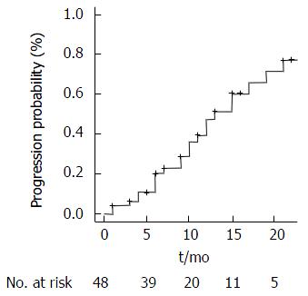
Figure 2 Time to progression.
- Citation: Greco G, Cascella T, Facciorusso A, Nani R, Lanocita R, Morosi C, Vaiani M, Calareso G, Greco FG, Ragnanese A, Bongini MA, Marchianò AV, Mazzaferro V, Spreafico C. Transarterial chemoembolization using 40 µm drug eluting beads for hepatocellular carcinoma. World J Radiol 2017; 9(5): 245-252
- URL: https://www.wjgnet.com/1949-8470/full/v9/i5/245.htm
- DOI: https://dx.doi.org/10.4329/wjr.v9.i5.245


