Copyright
©The Author(s) 2017.
World J Radiol. Apr 28, 2017; 9(4): 212-216
Published online Apr 28, 2017. doi: 10.4329/wjr.v9.i4.212
Published online Apr 28, 2017. doi: 10.4329/wjr.v9.i4.212
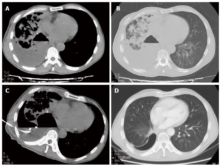
Figure 1 A 36-year-old male presented with fever and chest pain after right lung cancer resection.
A and B: Unenhanced transverse CT shows empyema in right pleura with liquid-gas plane, and infections can be seen in right lung; C: Catheater was placed under CT-guidance; D: Empyema and infections were absorbed after 2 mo.
- Citation: Li B, Liu C, Li Y, Yang HF, Du Y, Zhang C, Zheng HJ, Xu XX. Computed tomography-guided catheter drainage with urokinase and ozone in management of empyema. World J Radiol 2017; 9(4): 212-216
- URL: https://www.wjgnet.com/1949-8470/full/v9/i4/212.htm
- DOI: https://dx.doi.org/10.4329/wjr.v9.i4.212


