Copyright
©The Author(s) 2017.
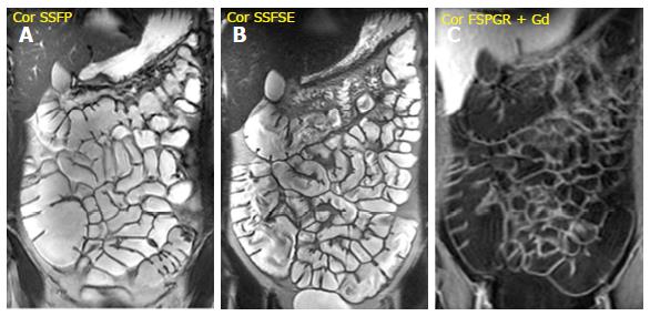
Figure 1 Assessment of small bowel anatomy, disease localization and segmental extension.
A: Steady-state free precession (SSFP); B: T2-weighted single shot fast spin echo (SSFSE) sequence; C: Gadolinium-enhanced fat-suppressed 3D spoiled gradient-echo (FSPGR) sequence.
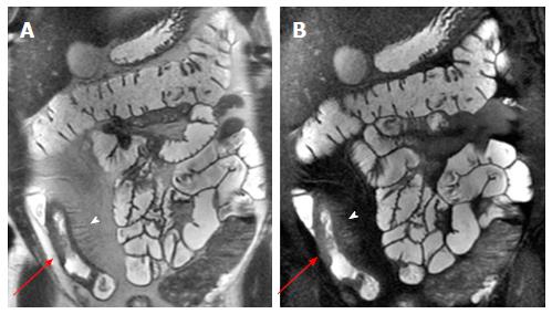
Figure 2 Wall thickening and mesenteric changes.
SSFSE (A) and gadolinium-enhanced FSPGR (B) sequences show wall thickening (red arrows) of terminal ileum with comb signs (arrowhead) and mesenteric fat proliferation. SSFSE: Single shot fast spin echo; FSPGR: Fat-suppressed 3D spoiled gradient-echo.
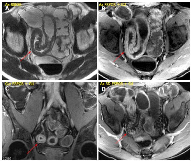
Figure 3 Active inflammation.
A: Wall thickening (10 mm) of terminal ileum extending for about 18 cm detected on SSFSE sequence; B: Gadolinium-enhanced FSPGR sequence shows the stratified enhancement pattern characterized by mucosal and muscle/serosa increased enhancement with intermediate hypointensity of edematous submucosa; C: Coronal FSPGR sequence revealing typical “target sign” due to stratified enhancement of bowel wall; D: Mesenteric fat thickening and vascular engorgement of vasa recta (comb sign) displayed on gadolinium-enhanced image. SSFSE: Single shot fast spin echo; FSPGR: Fat-suppressed 3D spoiled gradient-echo.
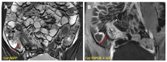
Figure 4 Subacute and stenotic disease with sinus tract.
A: SSFP sequence showing wall thickening (11 mm; red arrow) of terminal ileum with comb sign and mesenteric fat thickening; B: Post-gadolinium image reveals diffuse enhancement of the stenotic bowel loop and sinus tract (arrowhead), which is a blind-ending tract arising from the bowel wall. SSFP: Steady-state free precession; FSPGR: Fat-suppressed 3D spoiled gradient-echo.
Figure 5 Entero-vescical fistula.
A: Coronal SSFSE sequence detects wall thickening of the sigmoid colon with entero-vescical fistula (red arrow); B: FSPGR without gadolinium administration highlights the entero-vescical fistula, which appears hyperintense due to colonic content; C: Entero-vescical fistula appears as hyperintense transmural lines in post-gadolinium sequence. SSFSE: Single shot fast spin echo; FSPGR: Fat-suppressed 3D spoiled gradient-echo.
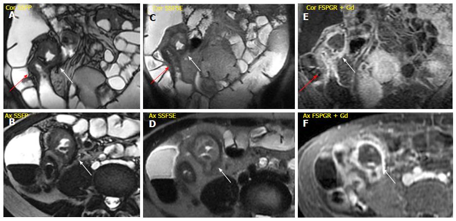
Figure 6 Peri-ileal abscess.
A-D: SSFP and SSFSE sequences display wall thickening of the terminal ileum associated (red arrow) with contiguous encapsulated collection of pus and inhomogeneous content (abscess, white arrow); E and F: FSPGR sequence shows mucosal enhancement with hypointense deep layers of the bowel wall (fibrotic disease), associated with enhancing peripheral rim of the capsulated collection (abscess, white arrow). SSFP: Steady-state free precession; SSFSE: Single shot fast spin echo; FSPGR: Fat-suppressed 3D spoiled gradient-echo.
Figure 7 Chronic disease.
A and B: Coronal SSFP and SSFSE sequences detect wall thickening (10 mm, red arrows) of neo-terminal ileum, after ileo-cecal resection, extending for about 19 cm; C: Coronal FSPGR sequence shows mucosal enhancement with hypointensity of the deep layers indicating the fibrotic disease. SSFP: Steady-state free precession; SSFSE: Single shot fast spin echo; FSPGR: Fat-suppressed 3D spoiled gradient-echo.
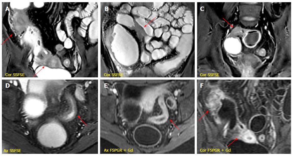
Figure 8 Fibrostenotic disease.
A-C: Multiple fibrotic strictures of the small bowel alternanting with prestenotic dilatated tracts detected on SSFSE sequences; D: Wall thickening of the sigmoid colon producing luminal narrowing displayed on SSFSE image; E and F: Post-gadolinium sequences reveal a diffuse and homogeneous enhancement in sigmoid colon (E) and small bowel (F) suggestive of subacute inflammation. SSFSE: Single shot fast spin echo; FSPGR: Fat-suppressed 3D spoiled gradient-echo.
- Citation: Mantarro A, Scalise P, Guidi E, Neri E. Magnetic resonance enterography in Crohn’s disease: How we do it and common imaging findings. World J Radiol 2017; 9(2): 46-54
- URL: https://www.wjgnet.com/1949-8470/full/v9/i2/46.htm
- DOI: https://dx.doi.org/10.4329/wjr.v9.i2.46


