Copyright
©The Author(s) 2016.
World J Radiol. Jun 28, 2016; 8(6): 600-609
Published online Jun 28, 2016. doi: 10.4329/wjr.v8.i6.600
Published online Jun 28, 2016. doi: 10.4329/wjr.v8.i6.600
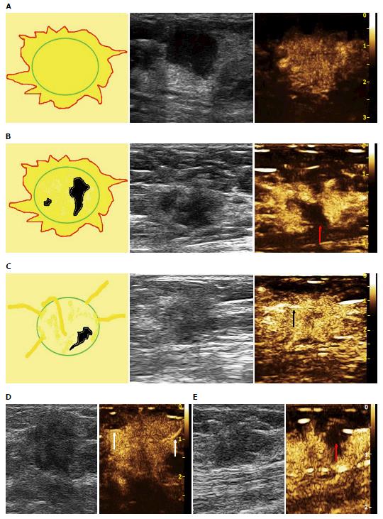
Figure 1 Predictive models for contrast-enhanced ultrasound of malignant breast lesions.
A: Hyper-enhancement, enlarged size after enhancement, irregular shape, IDC; B: Hyper-enhancement, centripetal enhancement, enlarged size, present perfusion defect (red arrow), IDC; C: Rapid wash-in with hyper-enhancement, present penetrating vessels (black arrow), IDC; D: Rapid wash-in with hyper-enhancement, present crab claw-like pattern (white arrows); IDC; E: Synchronous wash-in with iso-enhancement, with perfusion defect (red arrow), DCIS. IDC: Invasive ductal carcinoma; DCIS: Ductal carcinoma in suit.
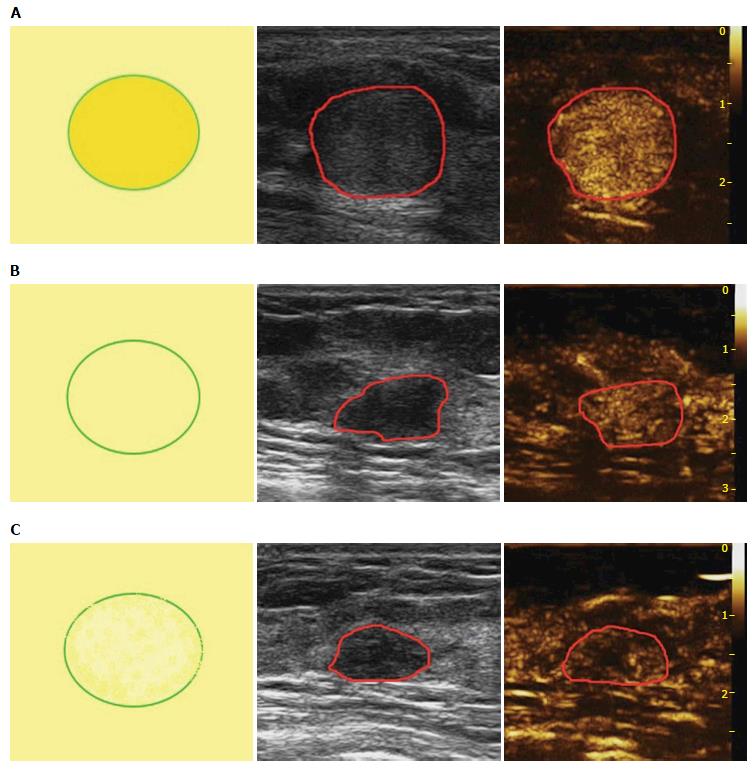
Figure 2 Predictive models for contrast-enhanced ultrasound of benign breast lesions.
A: Hyper-enhancement, equal size after enhancement, regular shape, without penetrating vessels, adenoma; B: Synchronous wash-in with iso-enhancement, can not distinguish margin and shape after enhancement, without perfusion defect or penetrating vessels, adenosis; C: Slow wash-in with hypo-enhancement, equal size after enhancement, without penetrating vessels, adenosis.
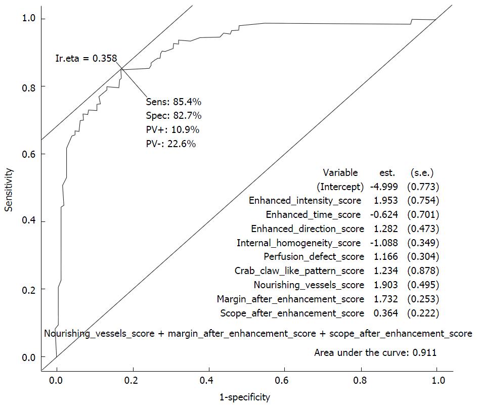
Figure 3 Receiver operating characteristic curve of logistic regression for contrast-enhanced ultrasound patterns.
Nine independent variables were identified in the final step of the logistic regression analysis forward model: Enhancement intensity, enhancement time, enhancement direction, internal homogeneity, perfusion defect, crab claw-like pattern, nourishing vessels, margin and scope after enhancement. Area under the curve was 0.911.
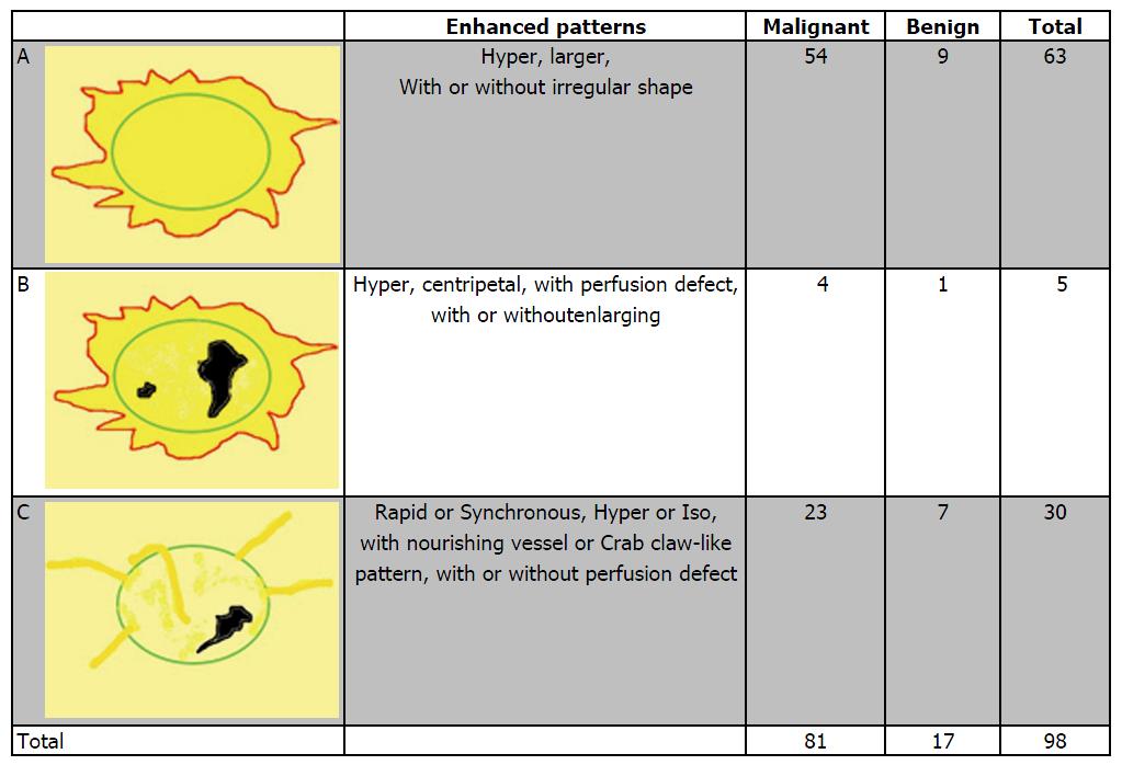
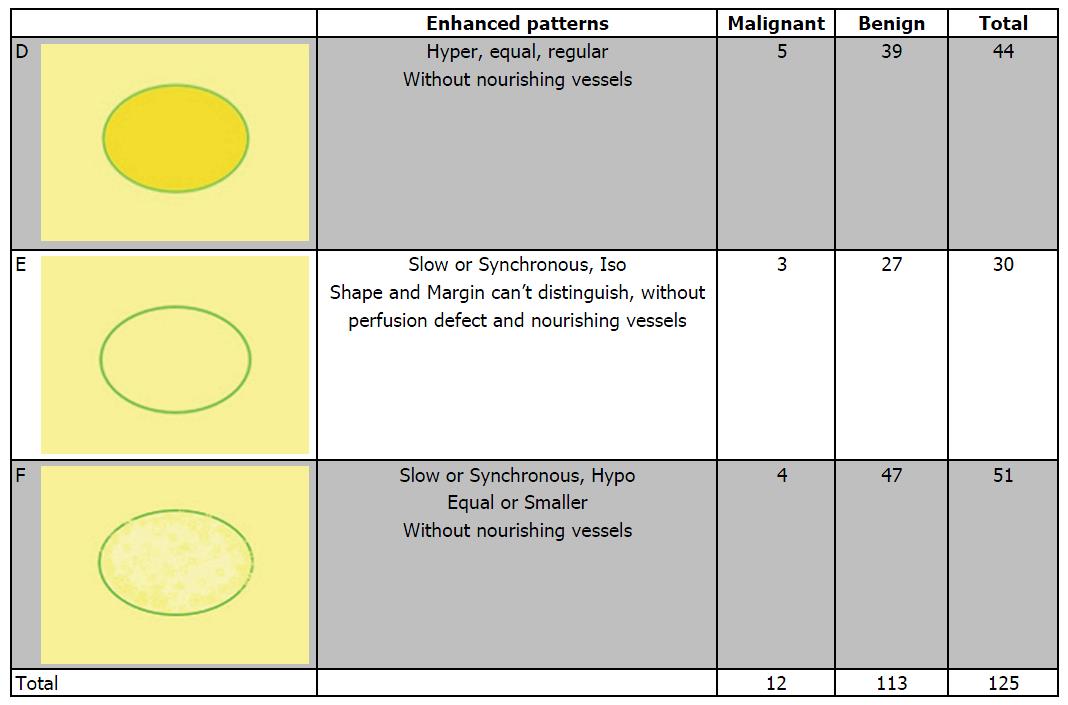
Figure 4 Distribution of pathologic results in different breast contrast-enhanced ultrasound malignant predictive models.
Figure 5 Distribution of pathologic results in different breast contrast-enhanced ultrasound benign predictive models.
- Citation: Luo J, Chen JD, Chen Q, Yue LX, Zhou G, Lan C, Li Y, Wu CH, Lu JQ. Predictive model for contrast-enhanced ultrasound of the breast: Is it feasible in malignant risk assessment of breast imaging reporting and data system 4 lesions? World J Radiol 2016; 8(6): 600-609
- URL: https://www.wjgnet.com/1949-8470/full/v8/i6/600.htm
- DOI: https://dx.doi.org/10.4329/wjr.v8.i6.600


