Copyright
©The Author(s) 2016.
World J Radiol. Jun 28, 2016; 8(6): 581-587
Published online Jun 28, 2016. doi: 10.4329/wjr.v8.i6.581
Published online Jun 28, 2016. doi: 10.4329/wjr.v8.i6.581
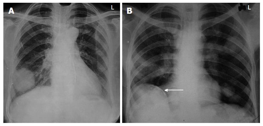
Figure 1 Uncomplicated hydatid cyst.
A: Posteroanterior view of chest X-ray showing well defined round radio-opacity in right lower zone; B: Chest X-ray showing multiple well defined round opacities in left lung. Also note presence of calcified cyst in liver (arrow in B), which makes diagnosis of hydatid cyst almost certain.
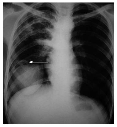
Figure 2 Air crescent sign: Chest X-ray showing well defined round radio-opacity in right lower zone with presence of a radiolucent rim at its superior aspect (arrow).
This sign is however not specific for hydatid cyst and can be seen in mycetoma, bronchogenic carcinoma, blood clot and pulmonary artery aneurysm.
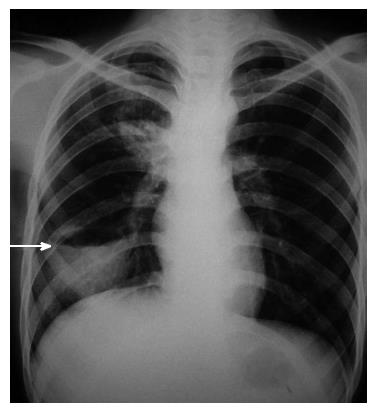
Figure 3 Complicated hydatid cyst: Chest radiograph of a patient who was a known case of hydatid cyst and presented with fever.
Air fluid level can be seen in the cyst (arrow) suggesting superimposed infection. Pyogenic lung abscess is the most common differential diagnosis for this radiological picture and computed tomography may be required to establish the diagnosis.
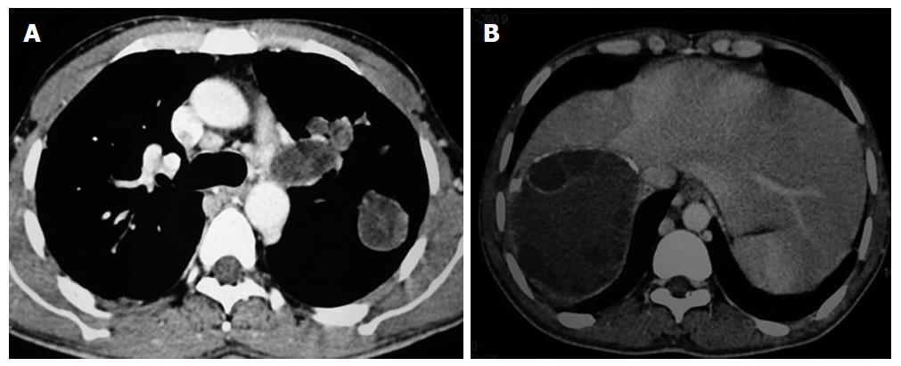
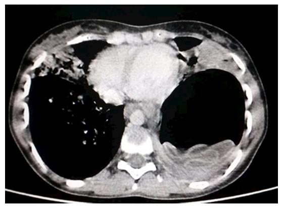
Figure 4 Hydatid cyst on computed tomography.
Axial contrast enhanced computed tomography showing multiple hydatid cysts in left lung (A) and liver (B) (same patient as in Figure 1B). Note peripheral calcification and daughter cysts in liver cyst.
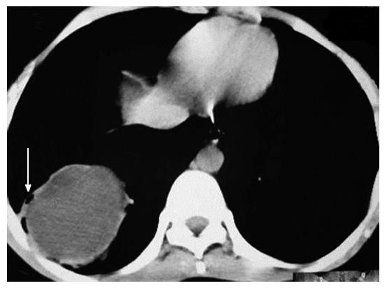
Figure 5 Axial contrast enhanced computed tomography images showing well defined cystic lesion with small air foci at periphery of the lesion (air bubble sign) (arrow).
Also note presence of mildly thickened wall with contrast enhancement (ring enhancement sign). This was a case of infected hydatid cyst.
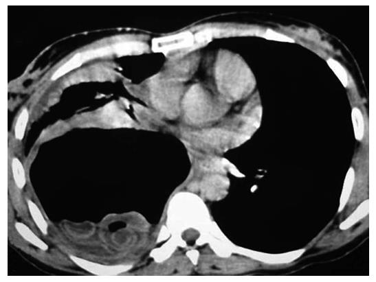
Figure 6 Axial computed tomography image in a case of ruptured hydatid cyst showing air and fluid with multiple curvilinear hyperattenuating membranes in dependant part (whirl sign).
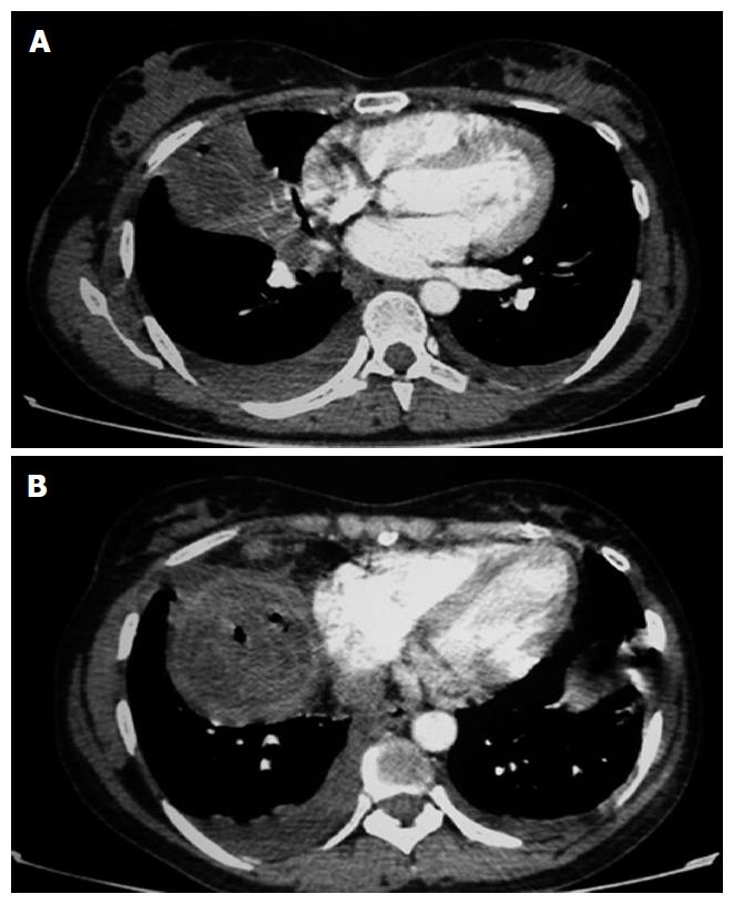
Figure 7 Complicated hydatid cyst showing ill defined wall with small air focus and consolidation in adjacent lung parenchyma (A) and posteroinferior to this cyst (B).
B showed air foci with serpingenous hyperattenuating membranes (“serpent sign”). Also note presence of mild bilateral pleural effusion, which was likely reactive.
Figure 8 “Water lily sign” on computed tomography.
Axial computed tomography image in a case of ruptured hydatid cyst showing air fluid level with crumpled endocyst appearing as floating membrane at air fluid interface.
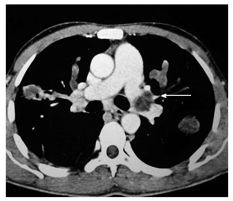
Figure 9 Axial contrast enhanced computed tomography showing hydatid cyst in left main pulmonary artery (arrow).
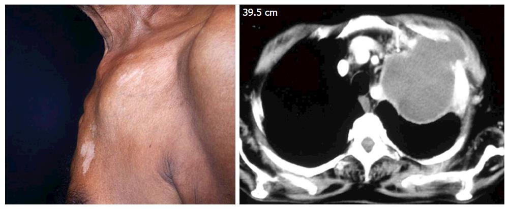
Figure 10 A 55-year-old male patient presented with swelling in left anterior chest wall.
Contrast enhanced computed tomography of the chest revealed hydatid cyst in left lung extending into left anterior chest wall.
- Citation: Garg MK, Sharma M, Gulati A, Gorsi U, Aggarwal AN, Agarwal R, Khandelwal N. Imaging in pulmonary hydatid cysts. World J Radiol 2016; 8(6): 581-587
- URL: https://www.wjgnet.com/1949-8470/full/v8/i6/581.htm
- DOI: https://dx.doi.org/10.4329/wjr.v8.i6.581


