Copyright
©The Author(s) 2016.
World J Radiol. Jun 28, 2016; 8(6): 537-555
Published online Jun 28, 2016. doi: 10.4329/wjr.v8.i6.537
Published online Jun 28, 2016. doi: 10.4329/wjr.v8.i6.537
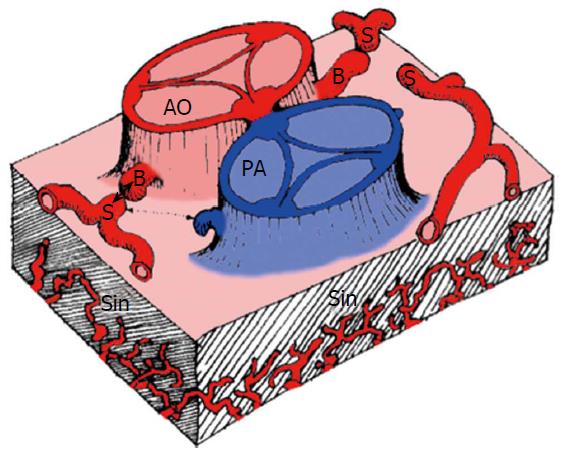
Figure 1 Schematic representation of the components involved in the embryogenesis of coronary arteries[3].
AO: Aorta; PA: Pulmonary artery; B: Coronary buds; S: Stems of right, circumflex and left anterior descending coronary artery; Sin: Sinusoids.
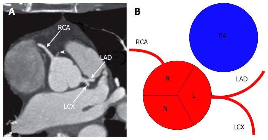
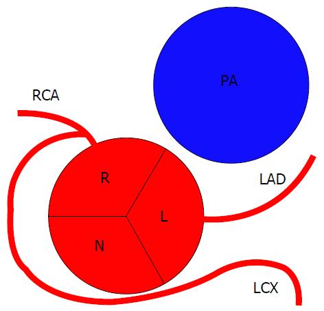
Figure 2 Conventional origin of coronary arteries.
A: Origin of coronary arteries on computed tomography angiography maximum intensity projection image, modified from[2]. Arrowhead shows a small conus branch arising from right sinus of Valsalva; B: Schematic representation of the origin of coronary arteries. RCA: Right coronary artery; LAD: Left anterior descending; LCX: Left circumflex artery; R: Right sinus of Valsalva; L: Left sinus of Valsalva; N: Non-coronary sinus; PA: Pulmonary artery.
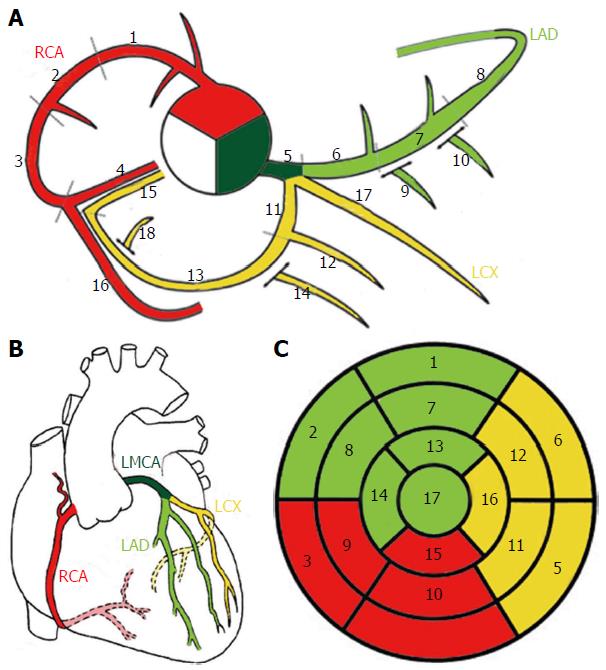
Figure 3 Schematic representation of normal coronary anatomy[11].
A: Coronary segments scheme proposed in SCCT guidelines for the interpretation and reporting of coronary computed tomography and used by Society of Cardiovascular Computer Tomography; B: Schematic representation of the coronary arteries. C: Circumferential polar plot of the 17 myocardial segments[56] with corresponding coronary artery territories. A-1, 2 and 3: Proximal, mid and distal RCA; 4: Right posterior descending artery; 5: Left main coronary artery; 6, 7 and 8: Proximal, mid and distal LAD; 9 and 10: First and second diagonal branches; 11: Proximal LCX; 12: First obtuse marginal branch; 13: Mid and distal LCX; 14: Second marginal branch; 15: Left posterior descending artery; 16: Right posterior-lateral branch; 17: Ramus intermedius; 18: Left posterior-lateral branch. RCA: Right coronary artery; LMCA: Left main coronary artery; LAD: Left anterior descending; LCX: Left circumflex artery.
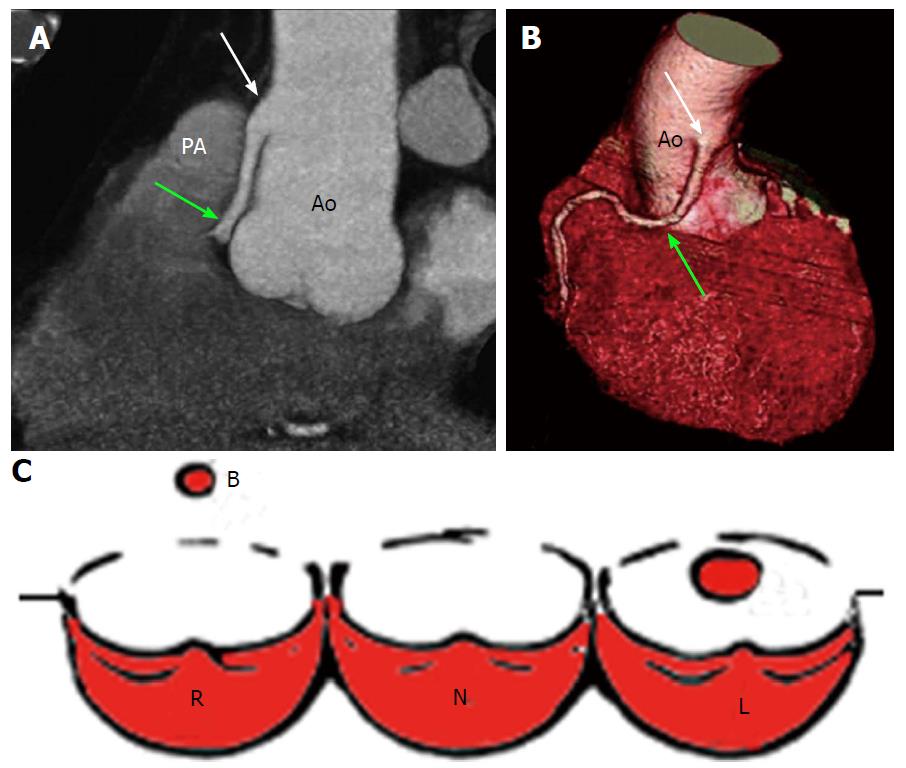
Figure 4 High take-off of right coronary artery in a 67-year-old man with equivocal exercise stress test[57].
Maximum intensity projection coronary CT angiogram (A) and three-dimensional volume-rendered CT reconstruction (B). Aortic step (white arrow) is seen to be the origin of RCA; RCA runs closely parallel to the proximal aorta (Ao) behind the pulmonary artery (PA). A: Regular position of left ostium; B: High position of right ostium; C: Schematic representation of high take-off. R: Right sinus of Valsalva; L: Left sinus of Valsalva; N: Non-coronary sinus; RCA: Right coronary artery; CT: Computed tomography.
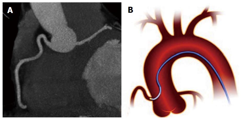
Figure 5 Shepherd’s crook right coronary artery.
A: From[14]; B: Schematic representation.
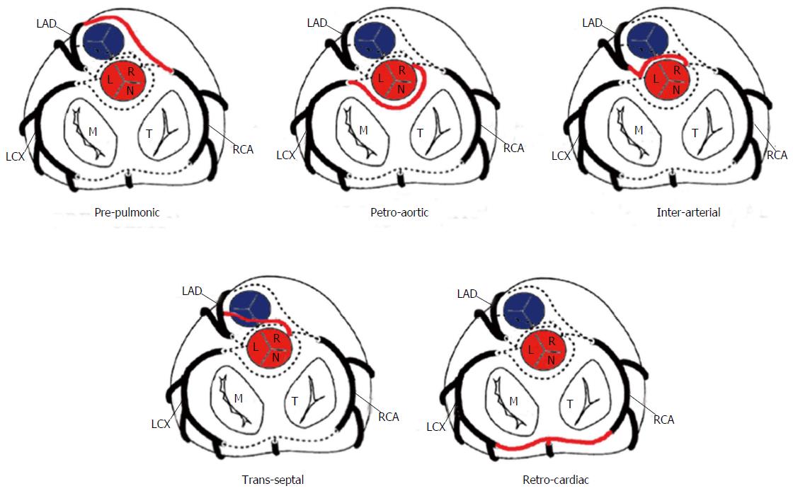
Figure 6 Schematic representation of the possible paths by which the coronary arteries can connect with the opposite coronary cusps[10].
LAD: Left anterior descending; LCX: Left circumflex; RCA: Right coronary artery; M: Mitral valve; T: Tricuspid valve; L: Left sinus of Valsalva; R: Right sinus of Valsalva; N: Non-coronary sinus.
Figure 7 Prepulmonic coronary artery in a 41-year-old man with chest pain[14].
CT image (A) and volume-rendered image (B) show a prepulmonic RCA (arrows) that courses in front of and just caudal to the pulmonary artery (PA) after arising from the LAD artery (arrowhead in A). On the right: Schematic representation of prepulmonic course. Ao: Aorta; LAD: Left anterior descending; LCX: Left circumflex; RCA: Right coronary artery; M: Mitral valve; T: Tricuspid valve; L: Left sinus of Valsalva; R: Right sinus of Valsalva; N: Non-coronary sinus.
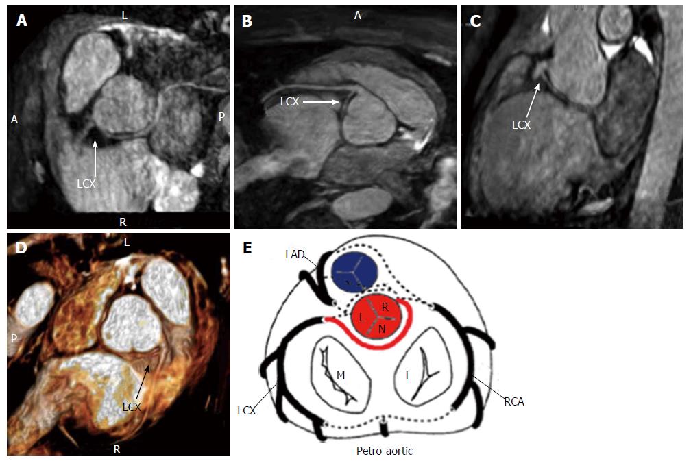
Figure 8 Anomalous origin of left circumflex from right coronary artery with retro-aortic course seen with three-dimensional cardiac magnetic resonance maximum intensity projection, multiplanar reformation and virtual reality imaging[19] (A-E).
E: Schematic representation of retro-aortic course. LAD: Left anterior descending; LCX: Left circumflex; RCA: Right coronary artery; M: Mitral valve; T: Tricuspid valve; L: Left sinus of Valsalva; R: Right sinus of Valsalva; N: Non-coronary sinus.
Figure 9 Example of interarterial course.
A and D: Anomalous origin of the right coronary artery from the left sinus of Valsalva with an interarterial course between the aorta and the pulmonary artery (arrow)[19]; B and C: Interarterial coronary artery in a 56-year-old man with chest pain. CT image (B) and volume-rendered image (C) show an interarterial LAD artery (arrow) arising from the RCA and coursing between the aorta (Ao) and pulmonary artery (PA). Note the short-segment narrowing of the proximal LAD artery, a finding likely related to the patient’s anomalous anatomy[14,15]; E: CT image, venous phase, three chamber view shows interarterial course of the left coronary artery between the aortic root and the right ventricular outflow tract (black arrow). RV: Right ventricle; LA: Left atrium; LV: Left ventricle; F: Schematic representation of interarterial course; LAD: Left anterior descending; LCX: Left circumflex; RCA: Right coronary artery; M: Mitral valve; T: Tricuspid valve; L: Left sinus of Valsalva; R: Right sinus of Valsalva; N: Non-coronary sinus.
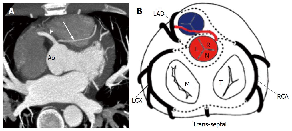
Figure 10 Trans-septal coronary artery in a 39-year-old man who underwent cardiac computed tomography after a stress test with equivocal results.
A: Axial computed tomography image shows a trans-septal LAD artery (arrow) that arises from the RCA (arrowhead)[14]; B: Schematic representation of trans-septal course. Ao: Aorta; RCA: Right coronary artery; LAD: Left anterior descending; LCX: Left circumflex; RCA: Right coronary artery; M: Mitral valve; T: Tricuspid valve; L: Left sinus of Valsalva; R: Right sinus of Valsalva; N: Non-coronary sinus.
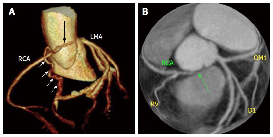
Figure 11 Cases of congenital atresia[16] (A and B).
Computed tomography revealed congenital atresia of RCA ostium, a hypoplastic proximal RCA that ends blindly (black arrow) and collaterals from left coronary system (white arrow). RCA: Right coronary artery; LMA: Left main coronary artery; RCA: Right coronary artery; D1: First diagonal branch, OM1: Obtuse marginal branch.
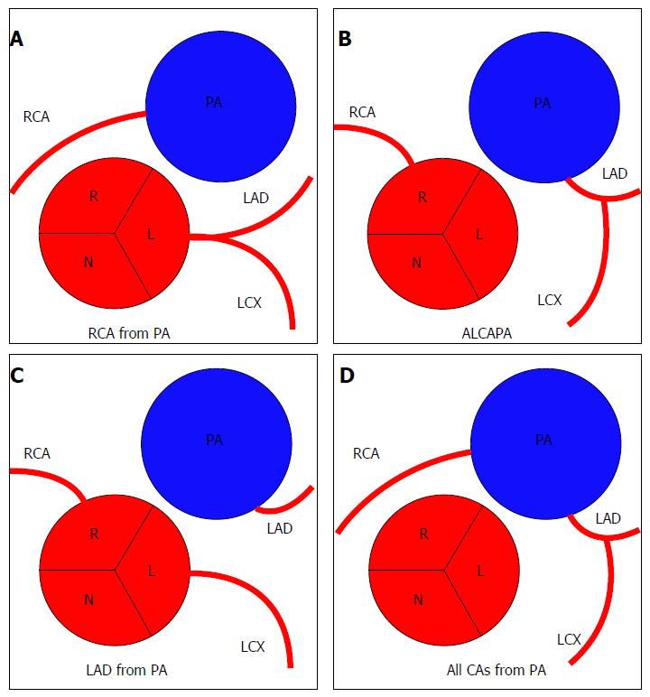
Figure 12 Anomalies of origin from pulmonary artery (A-D).
R: Right sinus of Valsalva; L: Left sinus of Valsalva; N: Non-coronary sinus; PA: Pulmonary artery; RCA: Right coronary artery; LAD: Left anterior descending; LCX: Left circumflex; ALCAPA: Anomalous left coronary artery from pulmonary artery; CAs: Coronary arteries.
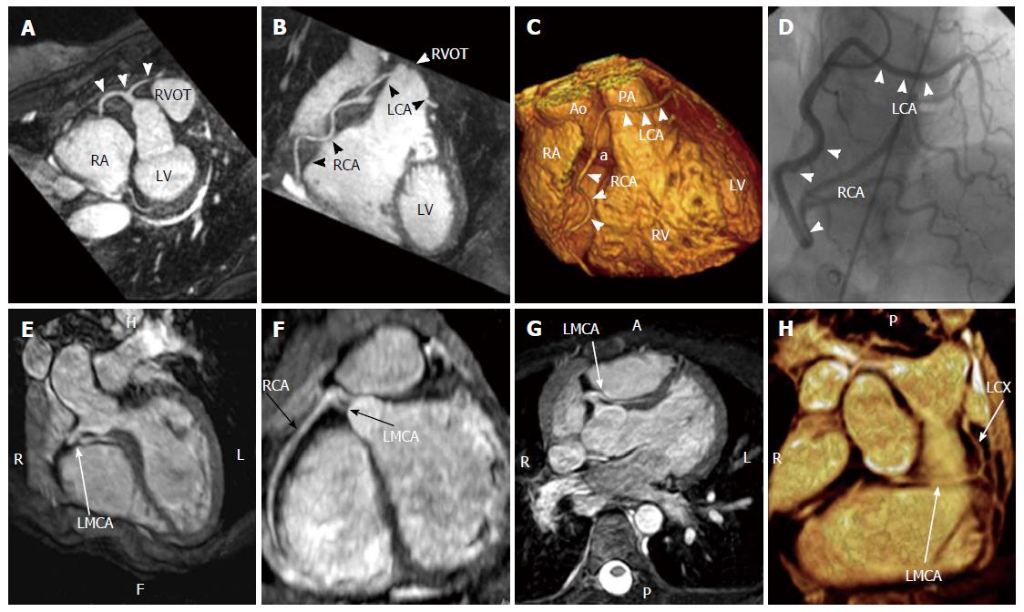
Figure 13 Cases of single coronary artery.
A-D: Coronary cardiac magnetic resonance, T2-prepared navigator-gated whole-heart technique shows the anomalous origin of a single coronary artery from the right sinus of Valsalva in a patient complaining of angina. A: Maximum intensity projection (MIP) image demonstrating the anomalous origin of the single coronary artery from the right sinus of Valsalva (white arrows); B: MIP image showing the path of the right coronary artery and of the left coronary artery, crossing in front of the right ventricle outflow tract; C: Volume rendering reconstruction (VRT) of the heart, showing the anomalous origin of the single coronary artery (a) from the aorta and the course of the RCA and the LCA; D: Coronary angiography in right anterior oblique projection, showing the anomalous coronary tree[17,18]; E-H: Case of single coronary artery arising from the right sinus of Valsalva with interarterial course of the left main coronary artery and distal origin of left circumflex (arrows) seen on three-dimensional cardiac magnetic resonance multiplanar reconstruction, MIP and volume rendering images[19]. Ao: Aorta; LCA: Left coronary artery; PA: Pulmonary artery; RVOT: Right ventricle outflow tract; RA: Right atrium; LMCA: Left main coronary artery; LCX: Left circumflex artery; RCA: Right coronary artery; LV: Left ventricle; RV: Right ventricle.
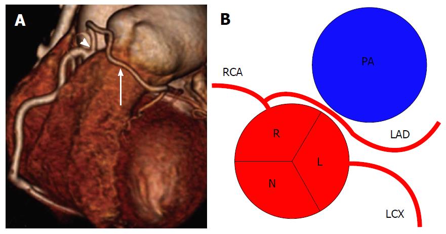
Figure 14 Left anterior desceding from right coronary artery[2].
A: Volume-rendered computed tomography image of LAD (arrow) artery arising from proximal RCA (arrowhead); B: Schematic representation. RCA: Right coronary artery; LCX: Left circumflex; LAD: Left anterior descending; R: Right sinus of Valsalva; L: Left sinus of Valsalva; N: Non-coronary sinus; PA: Pulmonary artery.
Figure 15 Schematic representation of left circumflex artery arising from right coronary artery.
RCA: Right coronary artery; LAD: Left anterior descending; LCX: Left circumflex; R: Right sinus of Valsalva; L: Left sinus of Valsalva; N: Non-coronary sinus; PA: pulmonary artery.
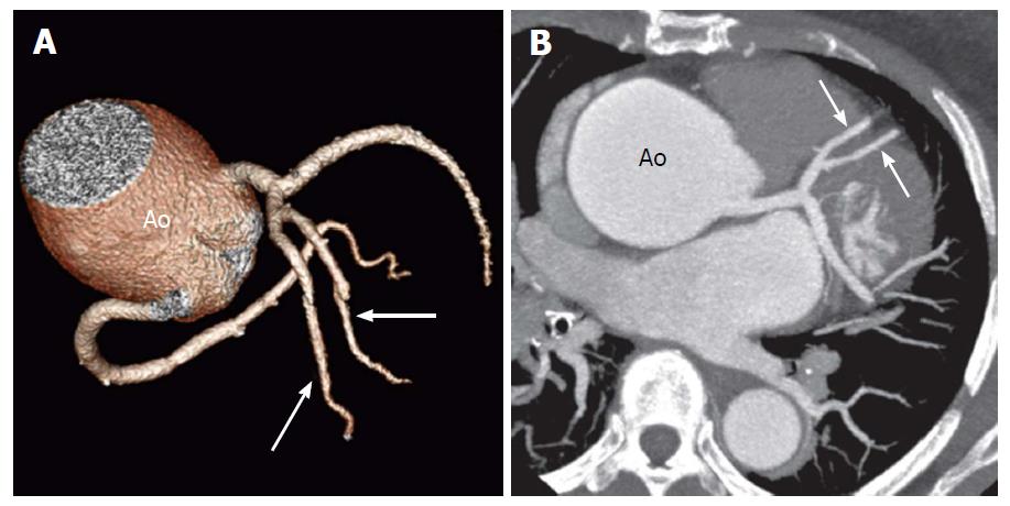
Figure 16 Duplication of the left anterior descending artery in a 52-year-old woman at preoperative evaluation[14].
Volume- rendered image (A) and oblique axial computed tomography image (B) show two equally sized LAD arteries (arrows). The Ao is noted to be aneurysmal. LAD: Left anterior descending; Ao: Aorta.
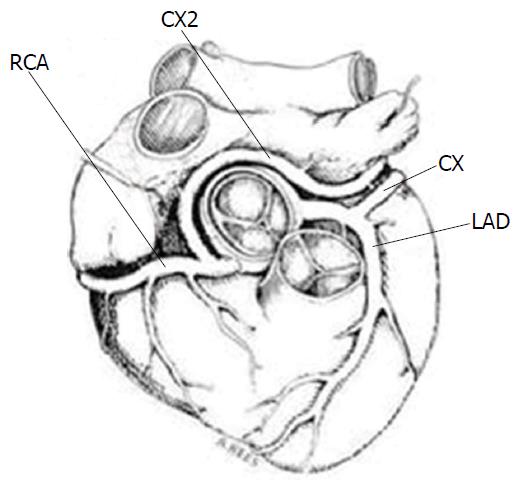
Figure 17 Schematic illustration of the double circumflex coronary system[4].
CX2 anomalous circumflex coronary artery arises from RCA. RCA: Right coronary artery; CX: Circumflex; LAD: Left anterior descending.
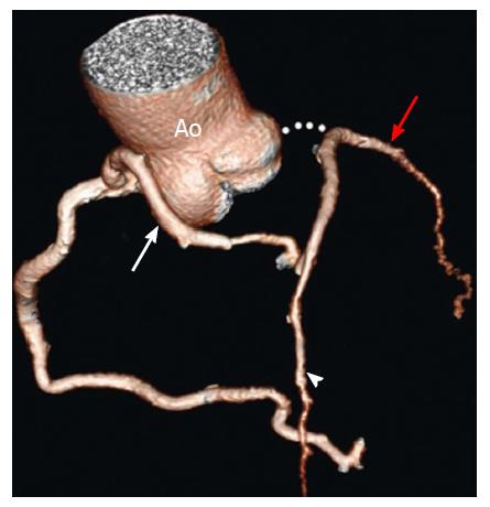
Figure 18 Atresia of the left main coronary artery in a 47-year-old woman with exertional chest pain and positive results on a stress test.
The patient presented with LMCA atresia in adulthood. Volume-rendered image shows large conus artery (white arrow) collateral to the left anterior descending artery (arrowhead). The LAD artery and LCX (red arrow) are diminutive overall[14]. LMCA: Left main coronary artery; LAD: Left anterior descending; LCX: Left circumflex; Ao: Aorta.
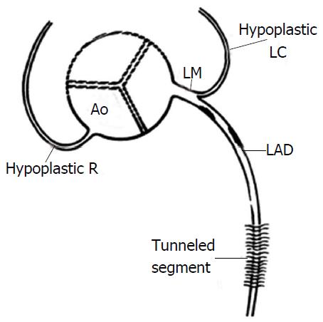
Figure 19 Congenital hypoplasia of right coronary artery and left circumflex artery[13].
LAD: Left anterior descending; Ao: Aorta; LM: Left main coronary artery; LC: Left circumflex; R: Right coronary artery.
- Citation: Villa AD, Sammut E, Nair A, Rajani R, Bonamini R, Chiribiri A. Coronary artery anomalies overview: The normal and the abnormal. World J Radiol 2016; 8(6): 537-555
- URL: https://www.wjgnet.com/1949-8470/full/v8/i6/537.htm
- DOI: https://dx.doi.org/10.4329/wjr.v8.i6.537


